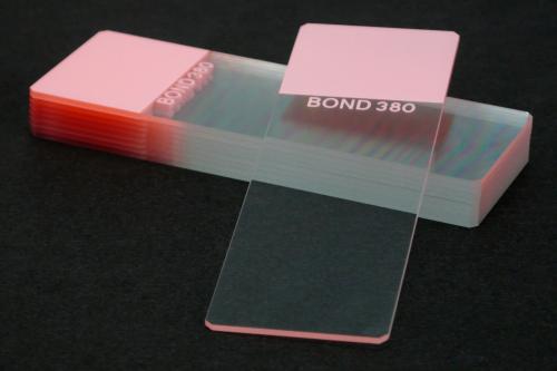
PAP Pen Liquid Blocker
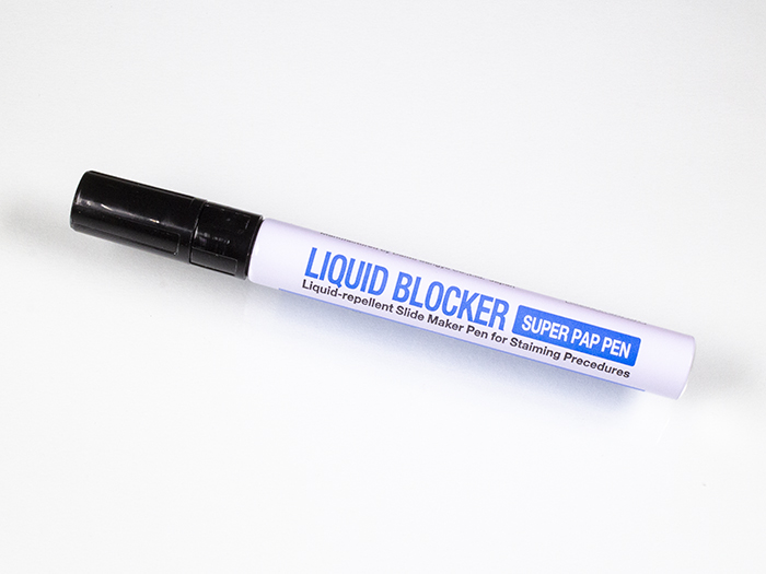
This newest generation of Pap Pens is still a superior hydrophobic liquid, which creates a secure boundary on glass and plastic around a tissue specimen during IHC procedures. Redesigned in 2001 for in-situ methods and stronger adherence. Fluid flows as a light blue on the slide and is easier to see than original (clear) pap pens.
Click here for information on the Mini Pap Pen Liquid Blocker
![]()

Newcomer Supply PAP Pen Liquid Blocker, hydrophobic slide markers for staining procedures, creates a visible hydrophobic barrier that provides proper surface tension to hold reagents within a targeted area when applied around tissue sections or smears on a microscopic slide. This surface tension ensures the amount of reagent needed for sufficient reaction is reduced and retained for complete tissue section coverage. Multiple specimens can be separated by drawn circles or lines applied to the same slide.
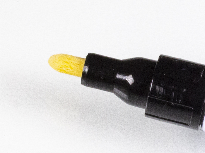
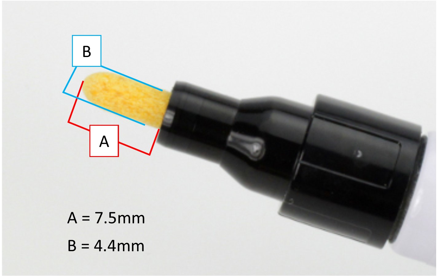
PAP Pen Liquid Blockers contain a unique formulation that is water repellent, insoluble in alcohol and acetone, soluble in xylene and temperature resistant up to 120°C. The PAP Pen Liquid Blocker, Mini provides a finer pen tip for drawing a thinner barrier film.
METHOD:
Technique: Paraffin, frozen sections and smears
- Manual staining for:
- Immunohistochemistry (IHC) procedures
- Immunofluorescence (IF) procedures
- In-Situ hybridization (ISH) procedures
- Enzyme procedures
PAP PEN ACTIVATION INSTRUCTIONS:
- Vigorously shake capped pen for approximately 10 seconds.
- Remove cap; hold pen upright and depress tip 2-3 times.
- Invert pen, press tip lightly on absorbent paper until tip is visually saturated.
- Release pressure on tip and blot away any excess liquid.
- Practice applying a thin PAP Pen liquid barrier on a test slide using light, minimal pressure. Applying too much pressure may result in excess release of barrier solution, creating a wider film that could touch tissue section edges.
- See Procedure Note #1.
- For optimum results; store PAP Pen Liquid Blocker at room temperature, tightly capped on a horizontal flat surface.
- Pen re-activation is not required unless tip dries out during storage.
PROCEDURE:
- Paraffin Section Method:
- Deparaffinize sections thoroughly in xylene and hydrate through 100% and 95% ethyl alcohols. Wash well with distilled water.
- Remove slide(s) from water or buffer; blot excess solution from slide and tissue section(s) with absorbent materials. Or place long edge of the slide on absorbent material to remove excess moisture.
- Encircle the tissue section(s) on the slightly damp slide surface with the PAP Pen. Do not touch the pen to any edges of the tissue section(s).
- See Procedure Note #2.
- Proceed to Step #10.
- Frozen Section and Smear Method:
- Apply PAP Pen barrier before fixation or immersion of slide(s) into water or buffer.
- Encircle the frozen tissue section(s) or smear on the slide with the PAP Pen. Do not touch the pen to any edges of the tissue section(s) or smear.
- See Procedure Note #2.
- After liquid barrier application, allow barrier film to dry before proceeding with staining procedure.
- See Procedure Note #3.
- Drain/rinse reagents off between staining steps; blotting slide and tissue as needed to remove excess solution.
- See Procedure Note #4.
- Complete staining process and coverslip with a compatible mounting medium. The barrier film will not affect the coverslipping procedure.
PROCEDURE NOTES:
- Once a tissue section is touched with PAP Pen liquid barrier it cannot be removed. The tissue section remains usable but colorization may result on the touched portion of tissue.
- If the tissue section is not completely encircled or segregated by the PAP Pen Liquid Blocker barrier film, reagents will not be fully retained on the tissue section and flood onto the slide. This may compromise adequate tissue coverage by reagents.
- If liquid barrier lines are not completely dry prior to staining, a precipitate from reaction with detection reagents may occur.
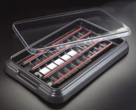
- The use of a Slide Moisture Chamber or StainTray™ (Part 68431, 68432, 6848 or 6847) is recommended for manual staining to maintain slide organization and a moist environment during the staining process.
REFERENCES:
- Grizzle, William, Cecil Stockard, and Paul Billings. “The Effects of Tissue Processing Variables Other Than Fixation on Histochemical Staining and Immunohistochemical Detection of Antigens.” The Journal of Histotechnology 24.3 (2001): 213-219.
- Vidwans, Malavika, Srinivas Mandavilli, Wanda Nethers, and Richard Cartun. “Fine-Needle Aspiration Diagnosis of a Neck Mass Using Immunocytochemical Stains Performed on Stained Cytology Slides.” The Journal of Histotechnology 25.4 (2002): 275-277.
- Modifications developed by Newcomer Supply Laboratory.