Background Buster
Eliminates non-specific binding in IHC, immunofluorescence, ELISA and flow cytometric assays. Allows staining of identical species antibodies and tissues (e.g. mouse antibody on mouse tissue). Short 10-20 minute incubation step prior to applying primary antibody. Shelf life 3 years. Store at 2-8°C.
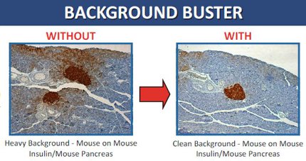
Innovex Background Buster is a peptide blocker that eradicates background staining for Mouse-On-Mouse IHC, ICC and immunofluorescence (IF), Human and Animal tissues IHC, ICC & IF staining in-situ Hybridization and Flow Cytometry and Immunoblotting. It is highly effective for quenching background fluorescence.
APPLICATION/INTENDED USE OF BACKGROUND BUSTER:
Background Buster is intended for eradicating non-specific binding and background in IHC, immunofluorescence labeling and in situ probe stains for both human and animal tissues.
BACKGROUND BUSTER FEATURES AND BENEFITS:
-
- Allows staining of identical species antibodies and tissue (e.g. mouse antibody on mouse tissue, rat-on-rat, rabbit-on-rabbit)
- Short 10-20 minute incubation step prior to applying primary antibody or in-situ probe at room temperature
- Delivers complete eradication of general background staining
- Replaces the use of normal serum, powdered milk, casein, and other blocking agents
- Excellent for both frozen and paraffin sections
STORAGE CONDITIONS:
Store in refrigerator at 2-8°C through the expiration date noted on the vial label.
PRODUCT FORMAT
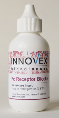
Working solution (Ready-to-Use). No dilution or adjustments required.
INSTRUCTIONS FOR USE:
Specimen Preparation for IHC Staining:
For paraffin sections: Deparaffinize sections and rehydrate in water.
For frozen sections: Cut sections, dry and fix in cold acetone or the fixative of choice. Incubate in PBS for 3 minutes at room temperature.
For cytocentrifuge preparation: Prepare cytocentrifuge preparations of cell suspensions and observe the following instructions:
- When using peroxidase enzyme conjugate label (staining with DAB or AEC), quench tissue endogenous peroxidase activity by immersing slides in 3% H2O2 in DI water and incubate for 10 minutes. Rinse with water.
- Apply 2-4 drops of Background Buster to achieve specimen coverage.
- Incubate for 10 minutes at room temperature for human tissues. For indirect species antibody and ANIMAL TISSUES, incubate for 20 minutes prior to the application of the primary antibody. For identical species tissue and antibody such as Mouse-on-Mouse, Mouse-on-Rat, Rat-on-Rat, incubate for 30 minutes prior to application of the primary antibody. For excessive general background staining or background staining due to endogenous biotin, incubate for 30 minutes.
- Rinse with water and proceed with IHC staining or immunofluorescence labeling or in-situ probe staining by following the manufacturer’s instruction.
For removal of endogenous biotin, Innovex Background Buster can be used for blocking endogenous biotin in place of avidin block or egg white. Tissues that are rich in biotin include kidney, liver and spleen.
- Apply 2-3 drops of Innovex Background Buster to achieve specimen coverage and incubate for 30 minutes at room temperature for both human and animal tissues prior to the application of the primary antibody.
- Rinse in water.
- Proceed with enzyme immunostaining or immunofluorescence or in-situ probe staining by following the manufacturer’s instruction.
Background Buster removes all background generated by cross-reactivity of primary antibodies with animal tissues.
- Apply 2-3 drops of Background Buster to achieve specimen coverage prior to the application of the primary antibody.
- For indirect species antibody and tissue such as Mouse-on-Rabbit, incubate for 20 minutes prior to the application of the primary antibody. For identical species tissue and antibody such as Mouse-on-Mouse, Mouse-on-Rat, Rat-on-Rat, incubate for 30 minutes prior to the application of the primary antibody.
- Proceed with immunostaining per staining kit instruction.
For in-situ stains: Apply Background Buster post hybridization and prior to the application of conjugated secondary antibody. Incubate for 10 minutes.
For Immunofluorescence labeling of tissues and cytosmears: Following the specimen preparation:
- Treat sections or smears with enough number of drops (3 to 6) of Background Buster to achieve specimen coverage.
- Incubate for 10-15 minutes at room temperature.
- Rinse in appropriate wash buffer and proceed with application of fluorochrome-conjugated antibody (direct method) or with the application of non-conjugated primary antibody, followed by fluorochrome conjugated secondary antibody (indirect method).
For Flow cytometric test samples: Test specimen consisting of blood cells or tumor cells suspension are treated as follows:
- Incubate cell suspensions with Background Buster in a test tube or in a microtiter plate with 0.2 ml/106 cells.
- Incubate for 5-10 minutes.
- Wash with the appropriate assay wash buffer and proceed with application of the conjugated (direct method) or unconjugated primary antibody followed by fluorochrome conjugated secondary antibody (indirect method).


