
Adhesive / Charged
The Right Adhesive Slide CAN make all the Difference!
So, which one is the right one?
A great question, but not an easy answer. There are some applications and staining platforms that specify a specific slide to use making the decision for you, but in most circumstances we will always recommend evaluating samples of the slide you’re interested in so that you can make the decision based on the actual results in your lab. Each lab may have their own unique way of doing things which may impact how an adhesive slide performs.
![]()
Application Categories with Adhesive Slide Recommendations:
|
Economical
|
General Purpose
(Histo & IHC) |
Histo & IHC Autostainers
|
Roche/Ventana Autostainers
|
|---|---|---|---|
| APS Poly-L-Lysine Premiere |
APS MAS-GP Superfrost Plus |
MAS-GP NewSilane Superfrost Plus |
(Manufacture Recommendation) Superfrost Plus TOMO |
|
Demanding IHC
|
Veterinary Animal Tissue
|
Difficult Tissue
|
Frozen Tissue
|
|
(With Antigen Retrieval)
|
(Dry, Fatty or Hard Tissue, such as bones, nails, breast, eyes & brain)
|
Charged vs. Coated Slides
You may hear techs referring to a charged or a coated slide, but keep in mind that both of these terms mean the same thing. A positive charged slide is referring to the positive charged arm that is anchored to the slide by a chemically coated surface. These coated surfaces could be poly-L-lysine, silane or some other proprietary surface chemistry.
Hydrophobic vs. Hydrophilic Slides
If you place a droplet of water onto a slide, you can quickly identify whether the slide is hydrophobic (water hating) or hydrophilic (water loving). If the water creates a tight bubble and doesn’t seem to freely run over the slide, you have a hydrophobic slide. On the other hand, if the water freely spreads out evenly, you have a hydrophilic slide.
There are definitely different degrees of hydrophobicity or hydrophilicity to slides, but in the extremes you will either have a tight, tall bubble or a completely covered slide with water.
Adhesive strength of a slide is independent of hydrophobic & hydrophilic characteristics
In trying to quickly assess a slide’s adhesive strength, one may mistakenly categorize hydrophobic adhesive slides as having stronger adhesion versus a hydrophilic adhesive slide. This would be an incorrect assumption due to the fact that the adhesive strength of a slide is independent to whether or not the slide has a hydrophilic or hydrophobic surface.
Advancements from adhesive slide manufactures from around the world have created proprietary surface chemistries that produce outstanding adhesion properties, while at the same time allowing them to create either a hydrophilic or hydrophobic surface for a given slide.
With hydrophobic surfaced adhesive slides, you will always get a faster interaction of the tissue section to the adhesive chemistry on that slide versus the same chemistry on a hydrophilic slide. Hydrophobic slides are quickly moving the water away from the surface of the glass, making the surface chemistry on the slide available to the tissue section almost immediately (speed is dependent on the level of hydrophobicity of the slide). Adhesive slides with a hydrophobic surface will make it much more difficult to refloat your tissue sections in the water bath versus a hydrophilic surfaced slide, which will allow you to continue to refloat and reposition your tissue sections in the water bath.
| TIPS |
Picking up tissue sections in a water bath with an adhesive slide
If using a hydrophobic surfaced slide, it is crucial to use good technique when picking up tissue sections in a water bath to minimize trapped water between the tissue section and the glass surface. Trapped water underneath tissue sections is much harder to remove with a hydrophobic surfaced slide versus a hydrophilic, due to the fact that tissue sections could form a sealed ‘bag’ like environment around the trapped water. If this occurs, folds may form in tissue sections after slide drying is complete. This can also have detrimental effects on antigen retrieval if the slide is being used in IHC protocols that require this procedure. The tissue section area that has folded over will be poorly adhered to the slide’s surface and is more susceptible to falling off when exposed to antigen retrieval steps. Hydrophilic surfaced slides will greatly reduce or eliminate the chance of trapped water underneath tissue sections when drying is taking place. This will enhance the tissue sections ability to lay flat on the slide minimizing wrinkles or having a tissue section area folding over on itself.
Drying slides vertically
If drying slides in a vertical position and it’s a high volume operation, one challenge that can arise when using a hydrophilic adhesive slide is tissue section migration. The tissue section may be perfectly placed on the slide, but after drying the tissue section is closer to the bottom of the slide. This happens when the water that is located between the tissue section and the slide’s surface is still present prior to the tissue section adhering to the adhesive slide’s surface chemistry. While the process of the water draining out between the two surfaces occur, the tissue section can move along with the water down the slide until the tissue section can interact directly with the slide’s surface chemistry.
A remedy for this could be to dry the slides in a flat and horizontal position, but this will take up a lot more surface space around your water bath(s), which could be limited. Another option would be to use a hydrophobic adhesive slide where the adhesion of the tissue section to the slide’s glass surface is more immediate. The reason for this is that the slide’s hydrophobic surface is quickly repelling the water away and allowing the tissue section to react and adhere to the adhesive chemistries that are on the slide’s surface, faster.
Autostainers
For most automated stainers, it is recommended to start with a neutral or hydrophilic slide. The reason for this recommendation is to minimize or prevent non-staining. If any of the solutions being delivered to a hydrophobic adhesive slide begins to puddle on the slide where there is no tissue section, it can create non-staining or poor mixing of the reagents being delivered. Neutral or hydrophilic slides will help to minimize or prevent this from happening.
Background staining on slides with silver stains and possible other problematic stains
As the adhesive strength becomes stronger, it can bind with some stains and create background staining on the glass. Silver stains, in general, have experienced this trade off. To reduce or minimize the background staining, a lab may need to modify the given staining procedure that is producing the problem (adjust temperature or timing for development of the stain).
Another issue that can arise is if a staining rack contains more slides than it’s designed to accommodate. This may limit or impede the movement of solutions on and in between slides causing unintended consequences. For instances, hematoxylin may create background staining on the adhesive slide surface, because the acid wash was ineffective.
Shelf Life and Storage of adhesive slides
To get the best performance out of adhesive slides, it is recommended to use them within a year of receipt. To help maximize the length of time the slides will perform at optimal levels, storage conditions are very important. Store them in a temperature and humidity controlled room or lab. Do not store directly on the floor. Keep the air tight packaging intact for individual boxes until time of use.
![]()
NewSilane Slides
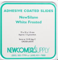
NewSilane Adhesive Microscope Slides
Adhesion: Very Strong
Application: Difficult and routine IHC. Difficult tissues with long incubations.
Unit: 72 slides/box, 1,440 slides/case
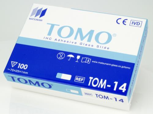
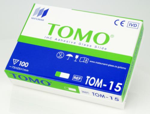
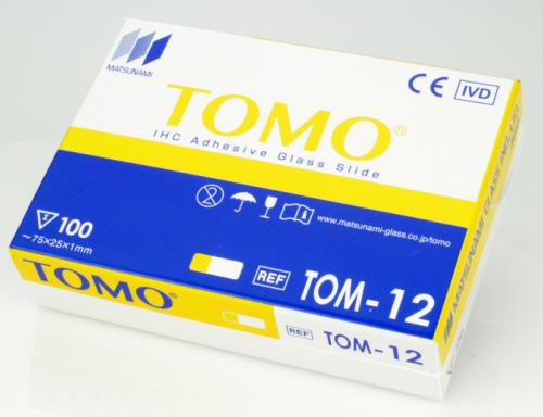
TOMO Slides
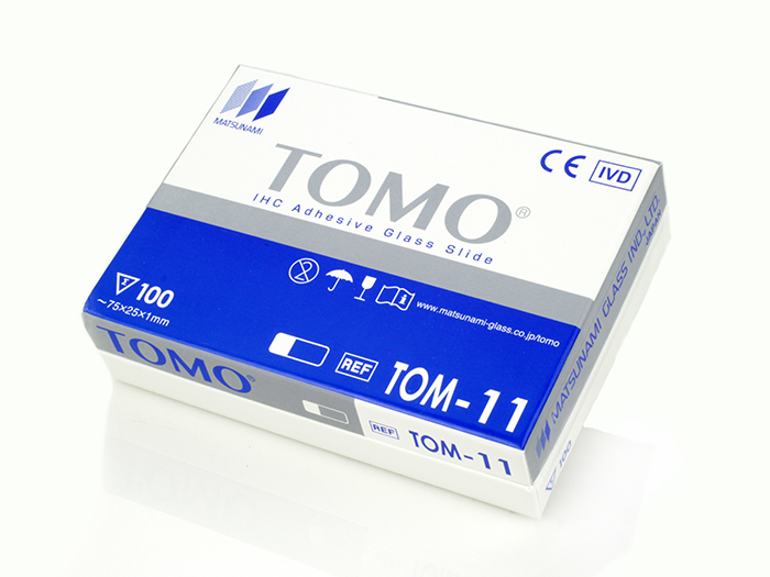
TOMO Adhesive Microscope Slides
Adhesion: Strong
Surface: Hydrophilic
Application: For automated IHC platforms such as Ventana/Roche Benchmark and Leica Bond
Unit: 100 slides/box, 1,000 slides/case


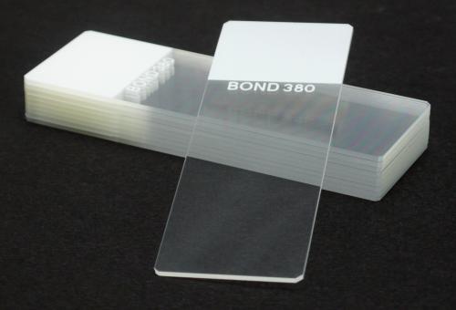
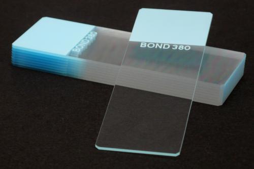
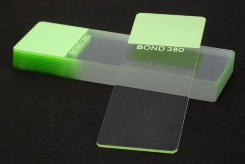
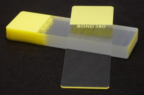
TruBond 380 Slides
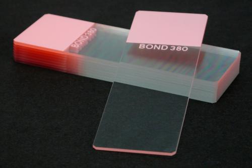
TruBond 380 Adhesive Microscope Slides
Adhesion: Very Strong
Surface: Hydrophilic
Application: For HIER processed when tissue type requires extra strong adhesion, epitope enhancement and DNA probe procedures
Unit: 100 slides/box, 1,000 slides/case


MAS Slides
MAS Adhesive Microscope Slides
Adhesion: Very Strong
Surface: Hydrophilic
Application: Excellent for the most challenging tissues and applications
Unit: 100 slides/box, 1,000 slides/case
MAS-GP Slides
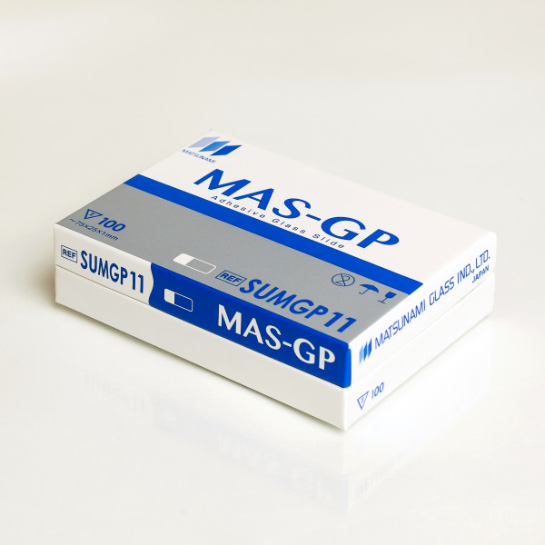
MAS-GP Adhesive Microscope Slides
Adhesion: Strong
Surface: Hydrophilic
Application: For all IHC applications, HIER, pH6/pH9 protocols, routine histology, and frozen sections
Unit: 100 slides/box, 1,000 slides/case
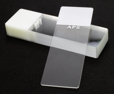
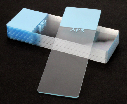
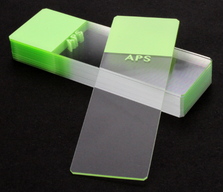
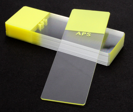
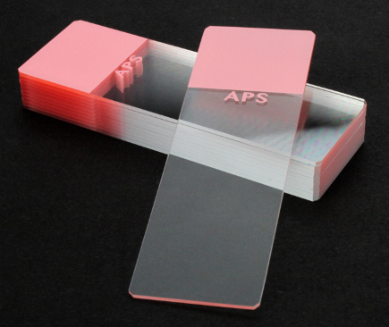
APS Slides
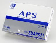
APS Adhesive Microscope Slides
Adhesion: Strong
Surface: Hydrophobic, treated to provide a permanent positive charge
Application: IHC and routine histology
Unit: 100 slides/box, 1,000 slides/case


Superfrost Plus Slides
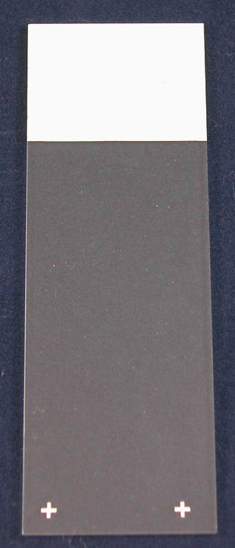
Superfrost Plus Adhesive Microscope Slides
-
-
- General purpose adhesive slide
- Dimensions: 75 x 25mm, 1mm thick
- Unit: Approx. 72 slides/box, 20 boxes/case
-
Premiere Slides
Premiere Charged Microscope Slides
-
-
- Aminosilane Coated Slides
- Imprint below colored end reads “Charged”
- Unit: 144 slides/gross, 10 gross/case
- Great pricing!
-
Aminosilane 75 x 50mm Slides
Aminosilane, 75 x 50 x 1.0 mm Frosted End
This slide offers a total of 55 x 50mm of an unobstructed adhesive surface for your tissue to lie down on.
-
- Dimensions: 75 x 50, 1 mm thick, “DOUBLE WIDE”
- Unit: Approx. 144 slides/gross
- 3/4″ Frosted End
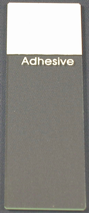
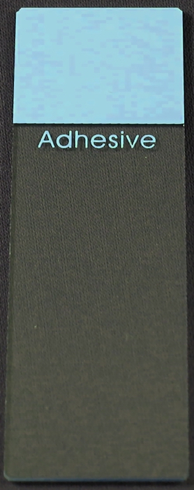
Poly-L-Lysine Slides

Poly-L-Lysine Adhesive Microscope Slides
Excellent for difficult tissues, frozen sections and special stains (especially Silvers).
-
-
- Imprint below colored end reads “Adhesive”
- Dimensions: 75 x 25 x 1 mm thick
- Unit: Approx. 144 slides/gross
-

| Part # 5010 | 1-9 gross (each) | $39.00 |
| Part # 5010 | 10-50 gross (each) | $36.50 |
| Part # 5010 | 51-99 gross (each) | $34.50 |
| Part # 5010 | 100+ gross (each) | Inquire |

| Part # 5013 | 1-9 gross (each) | $39.00 |
| Part # 5013 | 10-50 gross (each) | $36.50 |
| Part # 5013 | 51-99 gross (each) | $34.50 |
| Part # 5013 | 100+ gross (each) | Inquire |