Pigment, Substance & Mineral Stains
- Shelf Life for most Stain Kits is one year from date manufactured.
- Store Stain Kits at 15-30°C unless otherwise stated.
- Most Stain Kits include 2 Complimentary Positive Control Slides to be used for the initial verification of staining techniques and reagents.
- Stain solution groups are sold as individual products under separate part numbers with storage requirements and expiration date designated per bottle.
- H = Hazardous
For a more detailed chart regarding Pigments & Artifacts, click here.
| Name | Comments | Products | Part # |
| Copper | Some methods demonstrate copper associated protein rather than copper itself | Copper Stain Kit
Control Slides |
9113 |
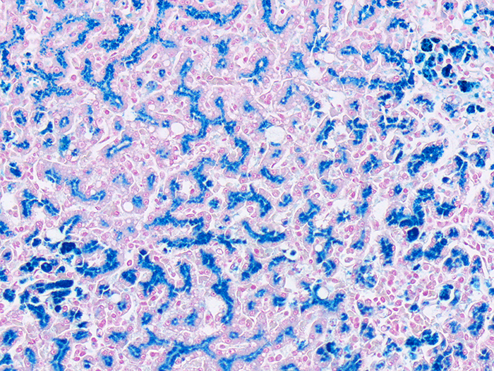
| Hemosiderin | Prussian Blue reaction stains ferric (+3) ions | Iron Stain Kit
Control Slides |
9136 |
| Hematoidin | Similar to bilirubin often formed as a result of hemorrhage, stains with bile methods but not with iron methods | ||
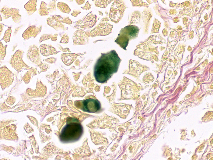
| Bile | Demonstrated when bilirubin is oxidized to biliverdin in an acid staining environment | Bilirubin Stain
Control Slides |
Procedure |
| Calcium | Black deposits formed when using silver reactions are due to reduction of silver by organic material followed by exposure to strong light | Calcium Stain
Control Slides |
Procedure |
| Urates |
Both forms are water soluble, aqueous fixatives should be avoided
Absolute ethanol is the fixative of choice
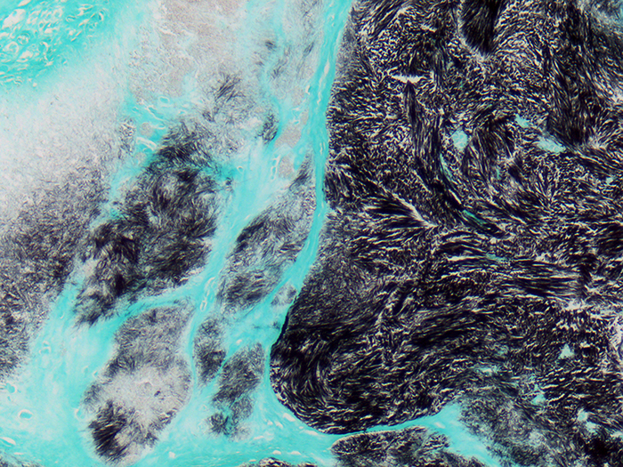
Gout – monosodium urate crystals appear yellow when their long axes are aligned parallel to a red compensator filter
Pseudogout -calcium pyrophosphate crystals, appear blue when their long axes are aligned parallel to a red compensator filter
|
Urates Stain
Control Slides |
Procedure |
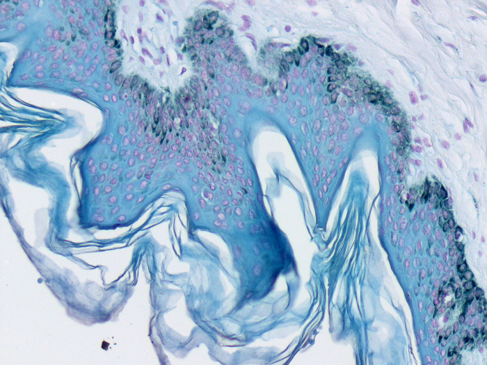
| Melanin | Identified by a number of methods can interfere with pathology interpretation if found in large amounts | Melanin Stain
Control Slides |
Procedure |
| Lipofusin | Stains variably but usually PAS positive | PAS Stain Kit | 9162 |
Artifact:
| Carbon | Inert and unreactive, resists removal procedures, commonly found in lung and mediastinal lymph nodes. | |
| Melanin | Can become problematic in large amounts and be removed by a variety of methods | Tech Memo |
| Formalin | Formed in unbuffered formalin when pH shifts to acidic, not reactive with iron staining methods | Tech Memo |
| Mercury | Deposited as a result of using mercury based fixatives | Tech Memo |
Amyloid, Bennhold Congo Red Stain Kit
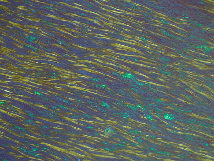
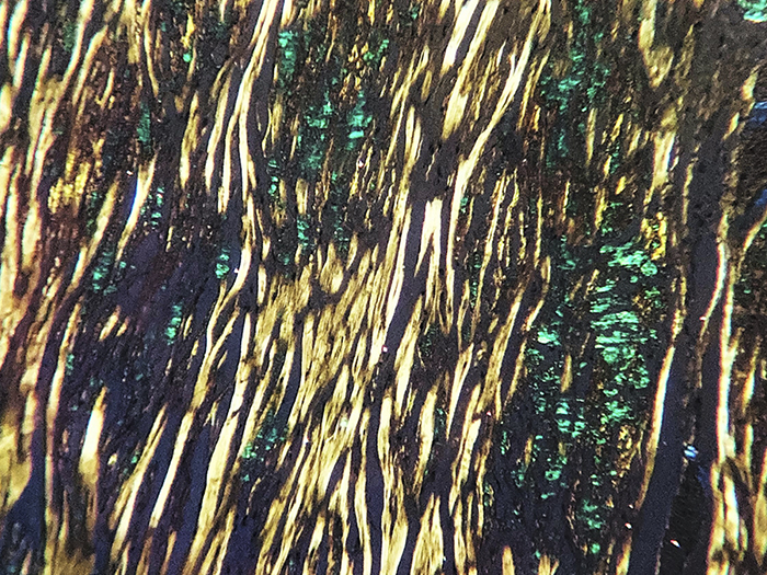
Identifies the extraneous protein deposits in amyloidosis. The use of polarizing lenses is the essential technique for visualizing amyloid positive areas and/or to confirm negativity.
-
-
- Microwave Modification Included
-
Amyloid, Puchtler Congo Red Stain Kit
Identifies the extraneous protein deposits in amyloidosis. The use of polarizing lenses is the essential technique for visualizing amyloid positive areas and/or to confirm negativity.


