Jones Basement Membrane Stain Kit
![]()
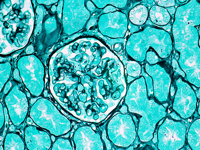
A straightforward silver technique for identification of glomerular and tubular basement membranes in renal tissue.
-
- No Titration!
![]()

JONES BASEMENT MEMBRANE STAIN KIT INCLUDES:
| Part 9167A | ||
| Solution A: | Methenamine 3%, Aqueous | 250 ml |
| Solution B: | Silver Nitrate 5%, Aqueous | 50 ml |
| Solution C: | Sodium Borate 5%, Aqueous | 50 ml |
| Solution D: | Periodic Acid 1%, Aqueous | 250 ml |
| Solution E: | Gold Chloride 0.25%, Aqueous | 250 ml |
| Solution F: | Sodium Thiosulfate 2.5%, Aqueous | 250 ml |
| Solution G: | Light Green SF Yellowish Stain 0.1%, Aqueous | 250 ml |
COMPLIMENTARY POSITIVE CONTROL SLIDES: Enclosed are two complimentary unstained positive control slides for the initial verification of staining techniques and reagents. Verification must be documented by running one Newcomer Supply complimentary positive control slide along with your current positive control slide for the first run. Retain the second complimentary control slide for further troubleshooting, if needed.
Individual stain solutions and additional control slides may be available for purchase under separate part numbers.
Additionally Needed:
| Hydrochloric Acid 5%, Aqueous | Part 12086 (for acid cleaning glassware) |
| Xylene, ACS | Part 1445 |
| Alcohol, Ethyl Denatured, 100% | Part 10841 |
| Alcohol, Ethyl Denatured, 95% | Part 10842 |
| Coplin Jar, Plastic | Part 5184 (for microwave modification) |
For storage requirements and expiration date refer to individual bottle labels.
APPLICATION:
Newcomer Supply Jones Basement Membrane Stain Kit procedure, with included microwave modification, is a silver technique for identification of glomerular and tubular basement membranes in renal tissue. A light green counterstain is used to enhance results.
METHOD:
Fixation: Formalin 10%, Phosphate Buffered (Part 1090)
Technique: Paraffin sections cut at 4 microns
Solutions: All solutions are manufactured by Newcomer Supply, Inc.
All Newcomer Supply Stain Kits are designed to be used with Coplin jars filled to 40 ml following the provided staining procedure. Some solutions in the kit may contain extra volumes.
PRESTAINING PREPARATION:
-
- If necessary, heat dry tissue sections/slides in oven.
- All glassware/plasticware must be acid cleaned prior to use.
-
- See Procedure Notes #1 and #2.
-
- Prepare Silver-Methenamine Working Solution and mix well:
-
- Solution A: Methenamine 3%, Aqueous 40 ml
- Solution B: Silver Nitrate 5%, Aqueous 2 ml
- Solution C: Sodium Borate 5%, Aqueous 4 ml
-
- Preheat Silver-Methenamine Working Solution to 45°-60°C in a water bath 20-30 minutes before use.
-
- Maintain solution between 45°-60°C to minimize precipitate.
- Do not preheat solution if using Microwave Modification.
-
STAINING PROCEDURE:
-
- Deparaffinize sections thoroughly in three changes of xylene, 3 minutes each. Hydrate through two changes each of 100% and 95% ethyl alcohols, 10 dips each. Wash well with distilled water.
-
- See Procedure Notes #3 and #4.
-
- Place in Solution D: Periodic Acid 1%, Aqueous for 15 minutes.
- Wash in tap water for 5 minutes; rinse in distilled water.
- Deparaffinize sections thoroughly in three changes of xylene, 3 minutes each. Hydrate through two changes each of 100% and 95% ethyl alcohols, 10 dips each. Wash well with distilled water.
-
- Incubate slides in preheated Silver-Methenamine Working Solution (Step #4) at 45°-60°C or at room temperature for 12-18 minutes until sections appear paper-bag brown.
- Periodically remove control, rinse in warm distilled water, check microscopically for adequate silver impregnation. Basement membranes should be dark brown. If tissue structures are not sufficiently dark, return slides to warm silver solution. Recheck at 2-3 minute intervals until desired intensity is achieved.
-
- Staining at room temperature will require longer incubation.
-
- Microwave Modification: See Procedure Note #5 .
-
- Place slides in a plastic Coplin jar with prepared Silver-Methenamine Working Solution (Step #3). Microwave at 70°C for 3 minutes.
- Check microscopically for adequate development.
- If additional incubation is required, return slides to heated silver solution and recheck at regular intervals.
-
- Rinse in three changes of distilled water.
- Tone in Solution E: Gold Chloride 0.25%, Aqueous for 1 minute.
- Rinse well in three changes of distilled water.
- Place in Solution F: Sodium Thiosulfate 2.5%, Aqueous; 2 minutes.
- Wash in tap water for 5 minutes; rinse in distilled water.
- Counterstain in Solution G: Light Green SF Yellowish Stain 0.1%, Aqueous for 1 minute.
- Quickly rinse slides in two changes of distilled water.
- Dehydrate in two changes each of 95% and 100% ethyl alcohol. Clear in three changes of xylene, 10 dips each; coverslip with compatible mounting medium.
RESULTS:
| Kidney glomerular basement membranes | Black |
| Intra-glomerular deposits | Black |
| Reticular fibers | Black |
| Nuclei | Outlined in black |
| Cytoplasm | Light green |
PROCEDURE NOTES:
-
- Acid clean all glassware/plasticware (Part 12086) and rinse thoroughly in several changes of distilled water.
- Plastic (Part 5500), plastic-tipped or paraffin coated metal forceps must be used with silver solutions to prevent precipitation of silver salts. No metals of any kind should come in contact with silver solutions. Only glass thermometers should be used.
- Drain slides after each step to prevent solution carry over.
- Do not allow sections to dry out at any point during procedure.
- The suggested microwave procedure has been tested at Newcomer Supply. This procedure is a guideline and techniques should be developed for use in your laboratory.
- If using a xylene substitute, closely follow the manufacturer’s recommendations for deparaffinization and clearing steps.
REFERENCES:
-
- Jones, David B. “Nephrotic Glomerulonephritis,” American Journal of Pathology2 (1957): 313–329.
- Luna, Lee G. Manual of Histologic Staining Methods of the Armed Forces Institute of Pathology. 3rd ed. New York: Blakiston Division, McGraw-Hill, 1968. 97-99.
- Sheehan, Dezna C., and Barbara B. Hrapchak. Theory and Practice of Histotechnology. 2nd ed. St. Louis: Mosby, 1980. 187-188.
- Modifications developed by Newcomer Supply Laboratory.