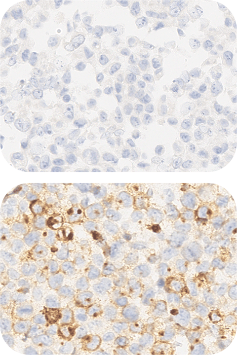
NTRK Analyte Control
HistoCyte Laboratories’ NTRK Analyte Control contains two cell lines that demonstrate positive and negative expression of NTRK. It specifically expresses wild type (WT) TrkA, that is recognized by the pan-NTRK antibodies as the WT Trk proteins and fusion proteins share a highly homologous c-terminus, within which the tyrosine kinase domain resides. Ideal for use as a same slide quality control in immunohistochemistry (IHC) to demonstrate the reagents have been applied to the slide. These cell lines are derived from the following tumors:
Cell line A: Breast adenocarcinoma
Cell line B: Large cell lymphoma
- Cells are fixed in 10% neutral buffered formalin and paraffin wax embedded.
- Sections are cut at 4µm, mounted on positively charged slides and dried overnight at 37ºC.
- Cell microarrays (CMA) contain cores that are 1.5-2mm in diameter and 3-3.5mm in length. It is possible to obtain over 300 sections depending on thickness.
- Not suitable for FISH as there is no translocation in the cell line. It is WT NTRK1.


