
Precision labeling for slides and cassettes—built for the demands of the histology lab
When every specimen matters, your labeling tool should never be the weak link. The HistoPlus™ Marking Pen is engineered specifically for histology workflows—delivering crisp, permanent markings that withstand the harsh chemicals, solvents, and processing conditions your samples encounter every day.
Why Choose HistoPlus™?
Confident Identification. Every Time.
HistoPlus™ pens produce bold, high-contrast markings that remain legible from grossing through staining and long-term storage. No smearing, fading, or ambiguity.
Engineered for Chemical Resistance
Designed to perform in real lab conditions, HistoPlus™ ink resists exposure to common histology reagents, including alcohols, xylene substitutes, and staining solutions—helping ensure your labels stay intact throughout processing.
Smooth, Controlled Application
A precision tip provides consistent ink flow for clean, accurate labeling on both slides and cassettes. Whether you’re writing fine identifiers or quick case numbers, you’ll get reliable performance without skipping.
Fast-Drying, Smear-Resistant Ink
Minimize handling errors and workflow interruptions. The quick-dry formula reduces smudging, even during high-throughput labeling.
Built for Daily Lab Use
Comfortable grip and durable construction make HistoPlus™ ideal for extended use at the bench, grossing station, or embedding area.
Key Features
-
- Permanent, solvent-resistant ink for histology applications
- Optimized for slides and cassettes (painted ends and plastic surfaces)
- Fine tip for precise, legible markings
- Fast-drying formula to reduce smearing
- Consistent ink flow—no skipping or blotting
- Reliable performance in demanding lab environments

CaviCide HP surface disinfectant offers a 1 minute universal contact time against 71 listed pathogens. Meeting all levels of the EPA’s Emerging Viral Pathogen Claim (EPA List Q), CaviCide HP utilizes a hydrogen peroxide formulation.
Features and Benefits:
-
- Efficient Disinfection: 1 step cleaning/disinfection
- Broad Spectrum Efficacy: Effective disinfection against 71 pathogens including MDRO’s (Multidrug Resistant Organisms)
- Ease of Use: Ready-to-Use liquid requires no mixing or dilution, reducing the risk of errors, saving time and improving compliance
- Low Toxicity Level: US EPA Toxicity Category IV product – No gloves needed!
Qualifications:
-
- Meets all levels of the EPA’s rigorous Emerging Viral Pathogen Claim (EPA List Q)
CaviCide HP List of Uses:
-
- Ambulance equipment surfaces
- Animal care facilities
- Bathrooms
- Correctional facilities
- Daycare centers
- Dental offices
- Emergency medical settings
- Emergency vehicles
- Exterior surfaces of anesthesia machines and respiratory therapy equipment
- Health club facilities
- Hospitals
- Infant/child care equipment surfaces
- Interior and exterior surfaces of infant incubators, bassinets
- Isolation areas
- Laboratories
- Laundry rooms
- Neonatal units
- Nursing homes
- Operating rooms
- Ophthalmic and optometric facilities
- Outpatient surgical centers
- Oxygen hoods
- Schools
- Surgical centers
CaviCide HP Kill Claims:
Mycobacterium
-
- Mycobacterium tuberculosis var: bovis (BCG)
Viruses (Non-Enveloped)
-
- Human Norovirus
- Human Rotavirus
- Rhinovirus Type 37
- Rhinovirus Type 1A
- Human Adenovirus Type 5
- Enterovirus Type D68
Viruses (Enveloped)
-
- Herpes simplex virus Type 1 & Type 2
- Avian Influenza A virus (H7N9)
- Avian Influenza A virus (H5N1)
- Influenza A virus (H3N2)
- Influenza B virus (H1N1)
- Parainfluenza virus Type 3
- Hepatitis B virus (HBV)
- Human Hepatitis C virus (HCV)
- Human lmmunodeficiency virus Type 1 (HIV-1)
- Severe Acute Respiratory Syndrome-related Coronavirus 2 (SARS-CoV-2)
- Vaccinia virus
- SARS-Associated Coronavirus (SARs-CoV)
- Cytomegalovirus (CMV)
- Influenza A virus (H1N1) Strains A/4108/2009 and A/04/09
- Human Coronavirus
- Respiratory Syncytial Virus (RSV)
- Measles virus
Pathogenic Fungi
-
- Trichophyton interdigitale
- Candida auris
- Candida albicans
- Ketoconazole-resistant Candida albicans
Bacteria
-
- Pseudomonas aeruginosa
- Staphylococcus aureus
- Salmonella enterica
- Bordetella pertussis
- Enterococcus faecium
- Klebsiella aerogenes
- Klebsiella pneumoniae
- Escherichia coli
- Escherichia coli O157:H7
- Burkholderia cepacia
- Stenotrophomonas maltophilia
- Streptococcus pyogenes
- Listeria monocytogenes
- Shigella dysenteriae
- Serratia marcescens
- Bordetella bronchiseptica
- Campylobacter jejuni
- Proteus vulgaris
Multi-Drug-Resistant Bacteria (MDRO)
-
- Penicillin-Resistant Streptococcus pneumoniae (PRSP)
- Vancomycin-Resistant Enterococcus faecalis (VRE)
- Methicillin-Resistant Staphylococcus aureus (MRSA)
- Hospital-Acquired and Community Acquired Methicillin-Resistant
Staphylococcus aureus (HA-MRSA and CA-MRSA) - Vancomycin Intermediate Staphylococcus aureus (VISA)
- Vancomycin Resistant Staphylococcus aureus (VRSA)
- Linezolid-Resistant Staphylococcus aureus (LRSA)
- Carbapenem-Resistant Klebsiella pneumoniae
- Multidrug-Resistant Acinetobacter baumannii (MRAB) Strains
ATCC 19606 and ATCC BAA-1605 - NDM-1 Enterobacter cloacae
- NDM-1 Klebsiella pneumoniae
- MBL Pseudomonas aeruginosa
- NMD-1 Escherichia coli
- ESBL Klebsiella pneumoniae
- ESBL Escherichia coli
- Multidrug-Resistant Klebsiella pneumoniae
- Carbapenem-Resistant Escherichia coli
- Carbapenem-Resistant Enterobacter cloacae
- Methicillin-Resistant Staphylococcus epidermidis (MRSE)
- Multi-Drug Resistant Pseudomonas aeruginosa
Also Available as CaviWipes HP – Click Here
![]()
PRODUCT SPECIFICATIONS:
Tissue: Positive staining gastrointestinal tract and negative staining lung.
Fixation: Formalin 10%, Phosphate Buffered (Part 1090).
Section/Glass: Paraffin sections cut at 4 microns on Superfrost™ Plus slides.
Quality Control Stain: Helicobacter quality control stained slide(s) included.
Reactivity: Guaranteed product specific reactivity for one year from date of receipt. Revalidate after one year to verify continued reactivity.
Storage: 15-30°C in a light deprived and humidity controlled environment.
Intended Use: To verify histological techniques and reagent reactivity.
Before using unstained control slides, review the enclosed stained slide(s) to ensure that this tissue source is acceptable for testing needs.
APPLICATION:
Newcomer Supply Helicobacter Control Slides are for the positive immunohistochemical staining of spiral shaped Helicobacter bacterium, strongly associated with inflammation of the stomach and implicated in the development of gastric malignancy, peptic ulcers, and chronic gastritis.
NEWCOMER SUPPLY VALIDATION PROCEDURE:
-
- Heat dry sections in oven according to your laboratory protocol.
- Deparaffinize sections thoroughly in three changes of xylene, 3 minutes each. Hydrate through two changes each of 100% and 95% ethyl alcohols, 10 dips each. Wash well with distilled water.
-
- See Procedure Note #1.
-
- Proceed with an epitope/antigen retrieval technique approved for use in your laboratory.
- Rinse in distilled water; tap off excess water.
- Circle sections with Pap Pen Liquid Blocker (Part 6505, 6506 or 6507) to reduce reagent usage and ensure tissue coverage.
- Block endogenous peroxidase with freshly made 3% Hydrogen Peroxide. Incubate for 5 minutes.
-
- See Procedure Note #2.
-
- Wash slides gently in distilled water. Rinse in two changes of Tris Buffered Saline.
-
- See Procedure Note #3.
-
- Tap off excess buffer; apply Helicobacter Pylori primary antibody. Incubate at room temperature for 30 minutes.
- Rinse slides in two changes of buffer.
- Tap off excess buffer; apply Amplifier. Incubate for 10 minutes.
- Rinse slides in two changes of buffer.
- Tap off excess buffer; apply HRP Polymer. Incubate for 10 minutes.
- Rinse slides in two changes of buffer.
- Prepare required quantity of DAB substrate/chromogen.
- Tap off excess buffer; apply DAB. Incubate for 5 minutes.
- Rinse slides in four changes of distilled water.
- Counterstain with Hematoxylin Stain, Gill I (Part 1180); 1-10 dips.
- Rinse slides in warm tap water to blue sections.
- Dehydrate in two changes each of 95% and 100% ethyl alcohol. Clear in three changes of xylene, 10 dips each; coverslip with compatible mounting medium.
RESULTS:
| Helicobacter sp. positive expression | Golden to dark brown bacteria |
| Lung | Negative |
| Nuclei | Blue |
PROCEDURE NOTES:
-
- Do not allow sections to dry out at any point during procedure.
- Dilute Hydrogen Peroxide 30%, Aqueous (Part 1206) with distilled water to a 3% (1/10) solution prior to use.
- Dilute Tris Buffered Saline 0.05M, pH 7.6, 10X (Part 140304) with distilled water to a 1/10 solution prior to use for all buffer rinses.
- Cell Marque Helicobacter pylori Polyclonal is the primary antibody used along with Cell Marque detection and ancillary reagents.
- If using a xylene substitute, follow manufacturer’s recommendation for deparaffinization and clearing steps.
REFERENCES:
-
- Cell Marque Helicobacter pylori Antibody datasheet.
- Modifications developed by Newcomer Supply Laboratory.
![]()
PRODUCT SPECIFICATIONS:
Tissue: Positive staining gastrointestinal tract.
Fixation: Formalin 10%, Phosphate Buffered (Part 1090).
Section/Glass: Paraffin sections cut at 4 microns on Superfrost™ Plus slides.
Quality Control Stain: Steiner-Steiner Modified quality control stained slide(s) included.
Reactivity: Guaranteed product specific reactivity for one year from date of receipt. Revalidate after one year to verify continued reactivity.
Storage: 15-30°C in a light deprived and humidity controlled environment.
Intended Use: To verify histological techniques and reagent reactivity.
Before using unstained control slides, review the enclosed stained slide(s) to ensure that this tissue source is acceptable for testing needs.
CONTROL SLIDE VALIDATION:
| With Steiner-Steiner Modified Silver Stain Kit: | Part 9171A | Individual Stain Solution | |
| Solution A: | Uranyl Nitrate 1%, Aqueous | 250 ml | Part 14036 |
| Solution B: | Silver Nitrate 1%, Aqueous | 250 ml | Part 13804 |
| Solution C: | Gum Mastic 2.5%, Alcoholic | 350 ml | Part 1145 |
| Ingredient D: | Hydroquinone, Powder | 5 grams | Part 12089 |
APPLICATION:
Newcomer Supply Helicobacter Control Slides are for the positive histochemical staining of helical shaped Helicobacter bacterium, associated with stomach inflammation and implicated in the development of gastric malignancy, peptic ulcers, and chronic gastritis.
PRESTAINING PREPARATION:
-
- Heat dry sections in oven according to your laboratory protocol.
- All glassware/plasticware must be acid cleaned prior to use.
-
- See Procedure Notes #1 and #2.
-
- Preheat Solution A: Uranyl Nitrate 1%, Aqueous to 60°C in a water bath. Save for Step #9.
- Preheat Solution B: Silver Nitrate 1%, Aqueous to 60°C in a water bath. Save for Step #11.
- Prepare Hydroquinone Solution; combine and mix well.
-
- Ingredient D: Hydroquinone, Powder 5 gm (or one rounded scoop with reusable mini sampling spoon)
- Distilled Water 25 ml
-
-
- Prepare fresh Reducing Solution by combining in order listed.
-
- Hydroquinone Solution (Step #5) 25 ml
- Solution C: Gum Mastic 2.5%, Alcoholic 15 ml
- Solution B: Silver Nitrate 1%, Aqueous 6 ml
- Solution will turn milky white after addition of Gum Mastic.
- Preheat solution in 45°C water bath. Save for Step #15.
-
- Do not preheat solutions if using Microwave Modifications.
- Prepare fresh Reducing Solution by combining in order listed.
STAINING PROCEDURE:
-
- Deparaffinize sections thoroughly in three changes of xylene, 3 minutes each. Hydrate through two changes each of 100% and 95% ethyl alcohols, 10 dips each. Wash well with distilled water.
-
- See Procedure Note #3
-
- Sensitize in preheated Solution A: Uranyl Nitrate 1%, Aqueous (Step #3) for 10 minutes in a 60°C water bath.
- Deparaffinize sections thoroughly in three changes of xylene, 3 minutes each. Hydrate through two changes each of 100% and 95% ethyl alcohols, 10 dips each. Wash well with distilled water.
Microwave Modification: See Procedure Note #4.
-
-
-
- Place slides in a plastic Coplin jar with Solution A: Uranyl Nitrate 1%, Aqueous. Microwave for 1 minute at 70°C.
-
- Rinse well in several changes of distilled water.
- Place slides in preheated Solution B: Silver Nitrate 1%, Aqueous (Step #4) and incubate in a 60°C water bath for 15 minutes.
-
Microwave Modification:
-
-
-
- Place slides in a plastic Coplin jar with Solution B: Silver Nitrate 1%, Aqueous. Microwave for 1 minute at 70°C.
- Remove from microwave, cover and let sit for 1 minute.
-
-
-
- Rinse well in several changes of distilled water.
-
- Excessive rinsing may cause nuclei to pick up silver.
-
- Rinse well in several changes of distilled water.
-
- Dip 5 times in two changes of fresh 95% and 100% ethyl alcohols.
- Place in Solution C: Gum Mastic 2.5%, Alcoholic for 3 minutes.
- Place slides in preheated Reducing Solution (Step #6) in 45°C water bath for 10-30 minutes with frequent agitation. Examine microscopically after 10 minutes of incubation.
-
- Check microscopically by dipping slide in 100% alcohol.
- Review for desired staining results.
- If necessary, return to warm solution; check every 2-5 minutes until desired results are achieved.
-
Microwave Modification:
-
-
-
- Place slides in a plastic Coplin jar with Reducing Solution. Microwave for 1 minute at 70°C. Remove from microwave.
- Pipette solution twice with plastic pipette to evenly distribute heated solution.
- Cover and let sit for 1 minute.
- Check microscopically by dipping slide in 100% alcohol.
- Review for desired staining results.
- If necessary, return to warm solution, check every 1 minute until desired results are achieved.
-
- Directly dehydrate in two changes of 100% ethyl alcohol. Clear in three changes of xylene, 10 dips each; coverslip with compatible mounting medium.
-
RESULTS:
| Helicobacter | Dark brown to black |
| Background | Golden brown |
PROCEDURE NOTES:
-
- Acid clean all glassware/plasticware (Part 12086) and rinse thoroughly in several changes of distilled water.
- No metals of any kind should come in contact with silver solutions to prevent precipitation of silver salts. Use plastic forceps (Part 5500) or paraffin coated metal forceps.
- Drain slides after each step to prevent solution carry over.
- The microwave procedure was tested using a laboratory-grade microwave oven. This procedure is a guideline and techniques should be developed for use in your laboratory.
- If using a xylene substitute, follow manufacturer’s recommendation for deparaffinization and clearing steps.
REFERENCES:
-
- Garvey, Winsome. “Some Favorite Silver Stains.” The Journal of Histotechnology 19.3 (1996): 269-278.
- Luna, Lee G. Histopathologic Methods and Color Atlas of Special Stains and Tissue Artifacts. Gaitheresburg, MD: American Histolabs, 1992. 218-219.
- Steiner, Gabriel and Grete Steiner. “New Simple Silver Stain for Demonstration of Bacteria, Spirochetes and Fungi in Sections of Paraffin Embedded Tissue Blocks.” Journal of Laboratory Clinical Medicine 29 (1944). 868-871.
- Swisher, Billie. “Modified Steiner Procedure for Microwave Staining of Spirochetes and Nonfilamentous Bacteria.” The Journal of Histotechnology 10.4 (1987): 241-243.
- Modifications developed by Newcomer Supply Laboratory.


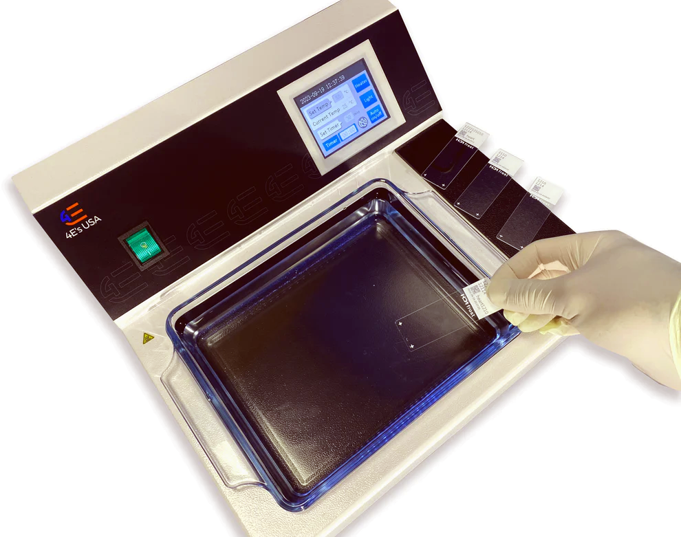
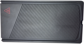
FEATURES OF THE SLIDE WARMER SDR200101:
-
- Digital control panel for precise temperature settings
- Working plate holds approximately 23 slides
- Durable, non-porous working plate surface
- Over-temperature protection
- Heater on indicator
- Alarm indicator
SPECIFICATIONS:
-
- Temperature range: Ambient to 75°C
- Temperature accuracy: ±1°C
- Power: 110w
- Overall dimensions: 10 1/4″ x 7 3/4″ x 3″
- Working plate dimensions: 9 3/4″ x 6 3/4″
- Slide capacity: Around 23 slides
- Weight: 5.3 lb.
WARRANTY DETAILS:
-
- Two year warranty
![]()
SOLUTION:
| 1 Gallon | |
| Decalcifying Solution, Formic/Formate | Part 10491C |
Additionally Needed:
| Decalcification End Point Set | Part 1051 |
For storage requirements and expiration date refer to individual bottle labels.
APPLICATION:
Newcomer Supply Decalcifying Solution, Formic/Formate (also known as Kristensen’s Decal) uses formic acid decalcifying agent along with an added formate buffer to help prevent cellular swelling and distortion during the decalcification process. This combination of reagents provides slow decalcification while maintaining excellent cellular morphology and is good for all bone specimen types and especially suitable for delicate bone samples.
METHOD:
Fixation: Formalin 10%, Phosphate Buffered (Part 1090)
-
-
- See Procedure Note #1.
-
Technique: Paraffin sections cut at 4 microns on adhesive slides
Solutions: All solutions are manufactured by Newcomer Supply, Inc.
PROCEDURE:
-
- Fix bone for a length of time sufficient for specimen size and type.
-
- See Procedure Note #2.
-
- Adequate bone fixation is essential before decal solution exposure.
- Wash fixed specimen in running tap water for 10 minutes.
- Submerge fixed bone segment in Decalcifying Solution, Formic/Formate, covering specimen at a 20:1 ratio.
-
- See Procedure Notes #3 and #4.
-
- Check the specimen regularly for sufficient solution coverage. Change solution daily and do not add or mix fresh solution with old.
- Decalcification with Decalcifying Solution, Formic/Formate can take 24 to 72 hours, dependent on specimen type, thickness and weight. Larger bones may require longer decal exposure.
- Check decal completion at regular intervals with Decalcification End Point Set (Part 1051) to deter over-decalcification.
-
- See Procedure Note #5.
-
- Wash in running tap water when decalcification is complete.
-
- Wash small samples 30-60 minutes.
- Wash larger bones 1-4 hours.
- Additional trimming of decalcified bone can occur at this point to size and thickness suitable for tissue processing.
-
- Proceed with tissue processing procedure for bone specimens.
- Trim block and section bone. If trimming or sectioning is impaired due to bone hardness, surface decalcification is recommended.
- Perform surface decalcification: Soak exposed bone surface side down in Decalcifying Solution, Formic/Formate for 15-60 minutes. Rinse block with distilled water to remove corrosive acids and re-section.
-
- See Procedure Note #6.
-
- Fix bone for a length of time sufficient for specimen size and type.
PROCEDURE NOTES:
-
- Other fixatives suitable for bone specimens include: AZF Fixative (Part 1009), B-5 Fixative Modified, Zinc Chloride (Part 1015), Bouin Fluid (Part 1020), Zamboni Fixative (Part 1459) and Zinc Formalin Fixative (Part 1482).
- Reduce size of a large bone by bisecting bone into smaller pieces, removing excess soft tissue for faster fixation. Maximum bone thickness of 3-5 mm is recommended.
- Decal solution should be in contact with all specimen surfaces. For multiple pieces, ensure pieces are separated or suspended and not in direct contact or stacked on each other.
- Enhance decal with low-speed agitation shaker, rotator or stir plate.
- Decalcification end-point testing can also be done with specimen radiography. Physical probing of bone is not recommended.
- Only a few calcium-free sections will be obtained after surface decalcification. Repeat the process for additional sections.
REFERENCES:
-
- Bancroft, John D. and Marilyn Gamble. Theory and Practice of Histological Techniques. 6th ed. Oxford: Churchill Livingstone Elsevier, 2008. 338-343.
- Luna, Lee G. Manual of Histologic Staining Methods of the Armed Forces Institute of Pathology. 3rd ed. New York: Blakiston Division, McGraw-Hill, 1968. 6-11.
- Ratcliffe, Norman A. “Some Useful Hints and Recipes for the Preparation and Examination of Stained Histological Sections.” In Practical Illustrated Histology. London: Palgrave Macmillan, 1982. 24-36.
- Urban, Ken. “Routine Decalcification of Bone.” Laboratory Medicine 12.4 (1981): 207-212.
- Villanueva, Anthony. “Experimental Studies in Demineralization and Its Effects on Cytology and Staining of Bone Marrow Cells.” The Journal of Histotechnology 9.3 (1986): 155-161.
- Modifications developed by Newcomer Supply Laboratory.


FEATURES OF THE PARAFFIN WAX DISPENSER:
-
- Insulated paraffin tank minimizes heat loss for energy efficiency but is cool to the touch for safety
- User friendly digital panel for precise temperature control and consistent results
- Consistent flow with a dedicated tap-heating system
- 0.5mm mesh filter screen delivers particle-free flow
- Rapidly melts 7.7 lb. of pellet wax in 45 minutes
- 3 position lever tap to control pouring
- Drip tray included for easy clean up
- Easily fill different container sizes with elevated tap
- Space saving with small footprint
SPECIFICATIONS:
-
- Temperature range: 0~100°C
- Accuracy: ±1°C
- Capacity: 7.5L
- Input Voltage: AC100V~120V, 50HZ
- Power: 1100W
- Dimensions: 17″ x 7″ x 17 3/4″
- Weight: 26.5 lb.
- Tap Height: 3 1/4″
- Tap Heating Intensity: Adjustable with the rotary know at the back
- Timer: 0-99:59 minutes (timer starts when it starts heating)
WARRANTY DETAILS:
-
- Two year warranty
Paraffin Wax Dispenser WXD101201 Operating Instructions


Enhance your histology experience with this advanced tissue flotation water bath, meticulously crafted for seamless operation. Digitally controlled for unparalleled ease, it boasts a longer and deeper water basin engineered for effortless handling of paraffin tissue sections.
Navigate effortlessly through its functionalities via the intuitive digital control panel. Illuminating your workflow, the glass basin is thoughtfully lit, ensuring optimal visibility for precise tissue placement.
FEATURES OF THE TISSUE FLOTATION BATH – DIGITAL:
-
- Extended-length glass dish
- Extra deep dish helps lift sections out of the bath easily
- Glass basin is lighted for easy tissue placement
- Digital control panel
- Precise heating capabilities with microprocessor controlled heating (Ambient to 75ºC)
- External temperature probe
- Temperature calibration
- Over heat protection
- Alarm indicator
SPECIFICATIONS:
-
- Temperature Range: RT-75°C
- Temperature Accuracy: ±1ºC
- Operational Temperature: 0-40°C
- Voltage: AC100-120V; AC200-240V; 50~60Hz
- Heating Power: 450 Watts
- Dimensions: 15″L x 13 3/4″W x 4 3/4″H
- Glass Basin Dimensions: 11 ½”L x 6 ½”W x 2″H
- Weight: 17.5 lb.
WARRANTY DETAILS:
-
- Two year warranty
Manual for Tissue Flotation Bath TFB100101
The KoolPlate™ cooling tray helps keep paraffin blocks chilled while preparing them for sectioning at the microtome and other laboratory procedures.
-
-
- Tray is made of thick wall high impact polystyrene
- Base is placed under the tray and made of Styrofoam
- Freeze tray for 16 hours in a conventional freezer
- Remains cold for 4 hours (when used with base)
- Trays are stackable
- Dimensions: 11 x 5 1/2 x 1
-
![]()
SOLUTION:
| Part 1047A | Part 1047B | Part 1047C | |
| Decolorizer, Aqueous | 500ml | 1 Liter | 1 Gallon |
Additionally Needed For H&E Staining:
| Hematoxylin and Eosin (H&E) Control Slides | Part 4278 |
| Xylene, ACS | Part 1445 |
| Alcohol, Ethyl Denatured, 100% | Part 10841 |
| Alcohol, Ethyl Denatured, 95% | Part 10842 |
| Hematoxylin Stain, Harris Modified | Part 1201 |
| Lithium Carbonate, Saturated Aqueous OR Scott Tap Water Substitute |
Part 12215 OR Part 1380 |
| Alcohol, Ethyl Denatured, 70% | Part 10844 |
| Eosin Y Working Solution OR Eosin Y-Phloxine B Working Solution |
Part 1072 OR Part 1083 |
For storage requirements and expiration date refer to individual product labels.
APPLICATION:
Newcomer Supply Decolorizer, Aqueous is a ready to use solution to improve staining contrast between nuclei and cytoplasm in automated regressive hematoxylin and eosin (H&E) stain protocols. Decolorizer, Aqueous works to slowly remove the correct amount of hematoxylin without concerns for over differentiation in automated H&E staining systems. For manual H&E staining, Decolorizer, Aqueous can be implemented for a more controlled differentiation step.
METHOD:
Fixation: Formalin 10%, Phosphate Buffered (Part 1090)
Technique: Paraffin sections cut at 4 microns
Solutions: All solutions are manufactured by Newcomer Supply, Inc.
H&E AUTOMATED STAINING WITH DECOLORIZER, AQUEOUS:
-
- If necessary, heat dry tissue sections/slides in oven or heat station.
- Deparaffinize and hydrate sections in the suggested number of stations and corresponding timings for xylenes and alcohols as specified per the automated stainer protocol.
- Wash well in distilled water.
- Stain with Hematoxylin Stain, Harris Modified (Part 1201), 2 to 5 minutes, depending on preference of nuclear stain intensity.
- Wash well in tap water.
- Differentiate in Decolorizer, Aqueous for 15 to 45 seconds.
-
- Timing depends on length of hematoxylin stain in Step #4.
- See procedure note #1.
-
- Rinse well in tap water.
- Blue in Lithium Carbonate, Saturated Aqueous (Part 12215) or Scott Tap Water Substitute (Part 1380) for 60 to 90 seconds.
- Wash well in tap water.
- Proceed to alcohol rinse for 1 minute.
-
- Use suggested percentage of alcohol per stainer protocol.
-
- Counterstain in Eosin Y Working Solution (Part 1072) or Eosin Y-Phloxine B Working Solution (Part 1083) for 30 seconds to 3 minutes, depending on preference of cytoplasmic stain intensity.
- Dehydrate and clear in the suggested number of stations and corresponding timings for alcohols and xylenes as specified per automated stainer protocol.
- Coverslip with compatible mounting medium.
RESULTS:
| Nuclei | Blue |
| Erythrocytes and eosinophilic granules | Bright pink to red |
| Cytoplasm and other tissue elements | Various shades of pink |
PROCEDURE NOTES:
-
- For manual H&E staining, follow regressive H&E stain protocol;
-
- Use Decolorizer, Aqueous in the hematoxylin differentiation step for 15 to 45 seconds.
- Timing will depend on length of hematoxylin stain exposure.
- Proceed through completion of the stain.
-
- If using a xylene substitute, follow manufacturer’s recommendation for deparaffinization and clearing steps.
- For manual H&E staining, follow regressive H&E stain protocol;
REFERENCES:
-
- Carson, Freida L. and Christa Cappellano. Histotechnology: A Self-Instructional Text. 5th ed. Chicago: ASCP Press, 2020. 122-125.
- Suvarna, S. Kim., Christopher Layton, and John D. Bancroft. Bancroft’s Theory and Practice of Histological Techniques. 8th ed. Oxford: Elsevier, 2019. 145-147.
- Modifications developed by Newcomer Supply Laboratory.
![]()
SOLUTION:
| Part 1046A | Part 1046B | Part 1046C | |
| Decolorizer, Alcoholic | 500 ml | 1 Liter | 1 Gallon |
Additionally Needed For H&E Staining:
| Hematoxylin and Eosin (H&E) Control Slides | Part 4278 |
| Xylene, ACS | Part 1445 |
| Alcohol, Ethyl Denatured, 100% | Part 10841 |
| Alcohol, Ethyl Denatured, 95% | Part 10842 |
| Hematoxylin Stain, Harris Modified | Part 1201 |
| Lithium Carbonate, Saturated Aqueous OR Scott Tap Water Substitute |
Part 12215 OR Part 1380 |
| Alcohol, Ethyl Denatured, 70% | Part 10844 |
| Eosin Y Working Solution OR Eosin Y-Phloxine B Working Solution |
Part 1072 OR Part 1083 |
For storage requirements and expiration date refer to individual product labels.
APPLICATION:
Newcomer Supply Decolorizer, Alcoholic is a ready to use solution to improve staining contrast between nuclei and cytoplasm in automated regressive hematoxylin and eosin (H&E) stain protocols. Decolorizer, Alcoholic works to slowly remove the correct amount of hematoxylin without concerns for over differentiation in automated H&E staining systems. For manual H&E staining, Decolorizer, Alcoholic can be implemented for a more controlled differentiation step.
METHOD:
Fixation: Formalin 10%, Phosphate Buffered (Part 1090)
Technique: Paraffin sections cut at 4 microns
Solutions: All solutions are manufactured by Newcomer Supply, Inc.
H&E AUTOMATED STAINING WITH DECOLORIZER, ALCOHOLIC:
-
- If necessary, heat dry tissue sections/slides in oven or heat station.
- Deparaffinize and hydrate sections in the suggested number of stations and corresponding timings for xylenes and alcohols as specified per the automated stainer protocol.
- Wash well in distilled water.
- Stain with Hematoxylin Stain, Harris Modified (Part 1201), 2 to 5 minutes, depending on preference of nuclear stain intensity.
- Wash well in tap water.
- Differentiate in Decolorizer, Alcoholic for 15 to 45 seconds.
-
- Timing depends on length of hematoxylin stain in Step #4.
- See procedure note #1.
-
- Rinse well in tap water.
- Blue in Lithium Carbonate, Saturated Aqueous (Part 12215) or Scott Tap Water Substitute (Part 1380) for 60 to 90 seconds.
- Wash well in tap water.
- Proceed to alcohol rinse for 1 minute.
-
- Use suggested percentage of alcohol per stainer protocol.
-
- Counterstain in Eosin Y Working Solution (Part 1072) or Eosin Y-Phloxine B Working Solution (Part 1083) for 30 seconds to 3 minutes, depending on preference of cytoplasmic stain intensity.
- Dehydrate and clear in the suggested number of stations and corresponding timings for alcohols and xylenes as specified per automated stainer protocol.
- Coverslip with compatible mounting medium.
RESULTS:
| Nuclei | Blue |
| Erythrocytes and eosinophilic granules | Bright pink to red |
| Cytoplasm and other tissue elements | Various shades of pink |
PROCEDURE NOTES:
-
- For manual H&E staining, follow regressive H&E stain protocol;
-
- Use Decolorizer, Alcoholic in the hematoxylin differentiation step for 15 to 45 seconds.
- Timing will depend on length of hematoxylin stain exposure.
- Proceed through completion of the stain.
-
- If using a xylene substitute, follow manufacturer’s recommendation for deparaffinization and clearing steps.
- For manual H&E staining, follow regressive H&E stain protocol;
REFERENCES:
-
- Carson, Freida L. and Christa Cappellano. Histotechnology: A Self-Instructional Text. 5th ed. Chicago: ASCP Press, 2020. 122-125.
- Suvarna, S. Kim., Christopher Layton, and John D. Bancroft. Bancroft’s Theory and Practice of Histological Techniques. 8th ed. Oxford: Elsevier, 2019. 145-147.
- Modifications developed by Newcomer Supply Laboratory.
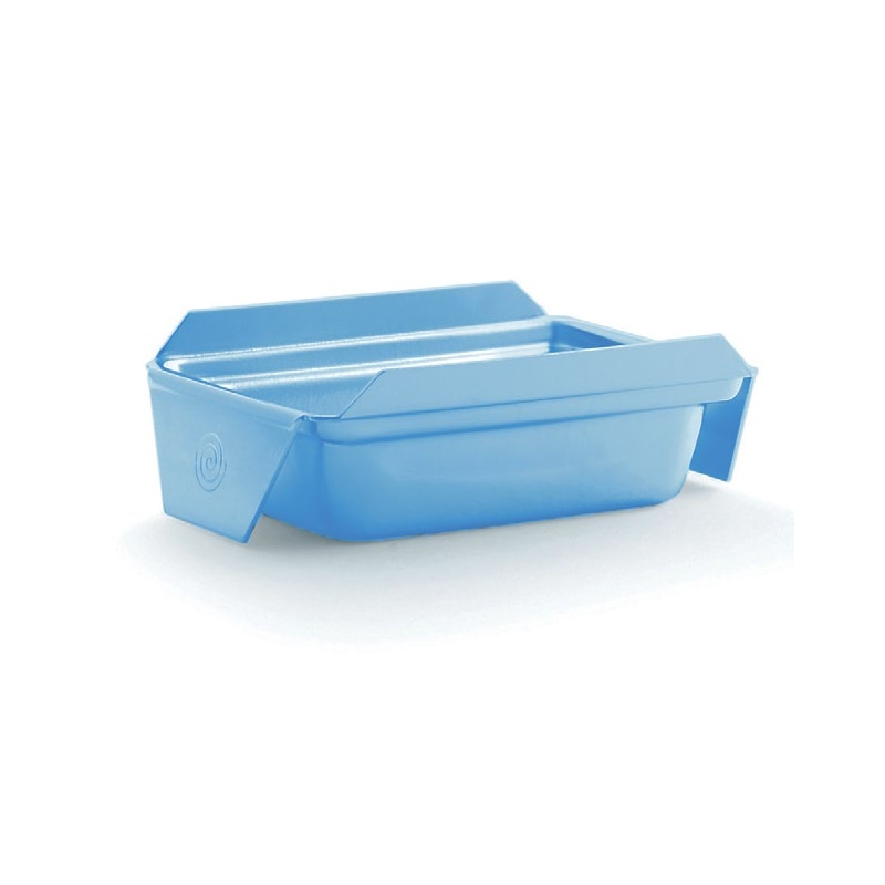
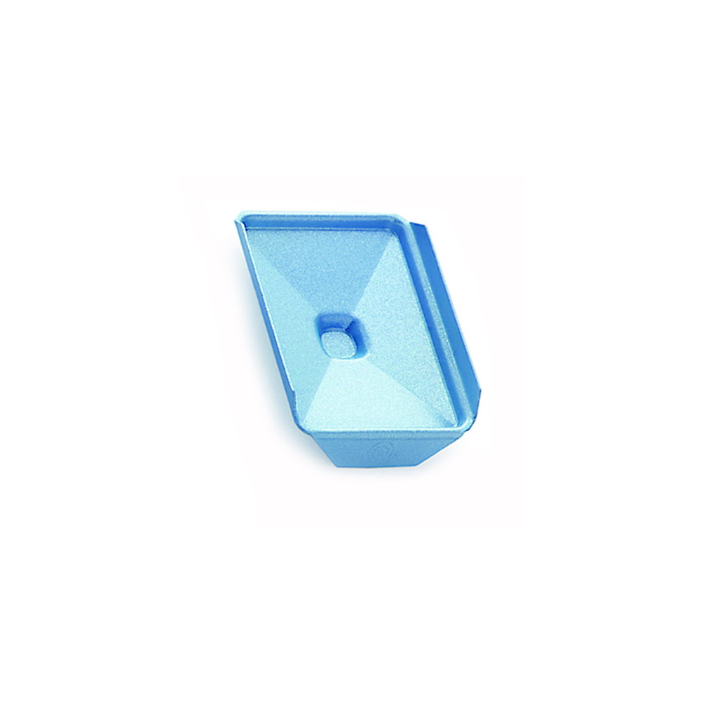
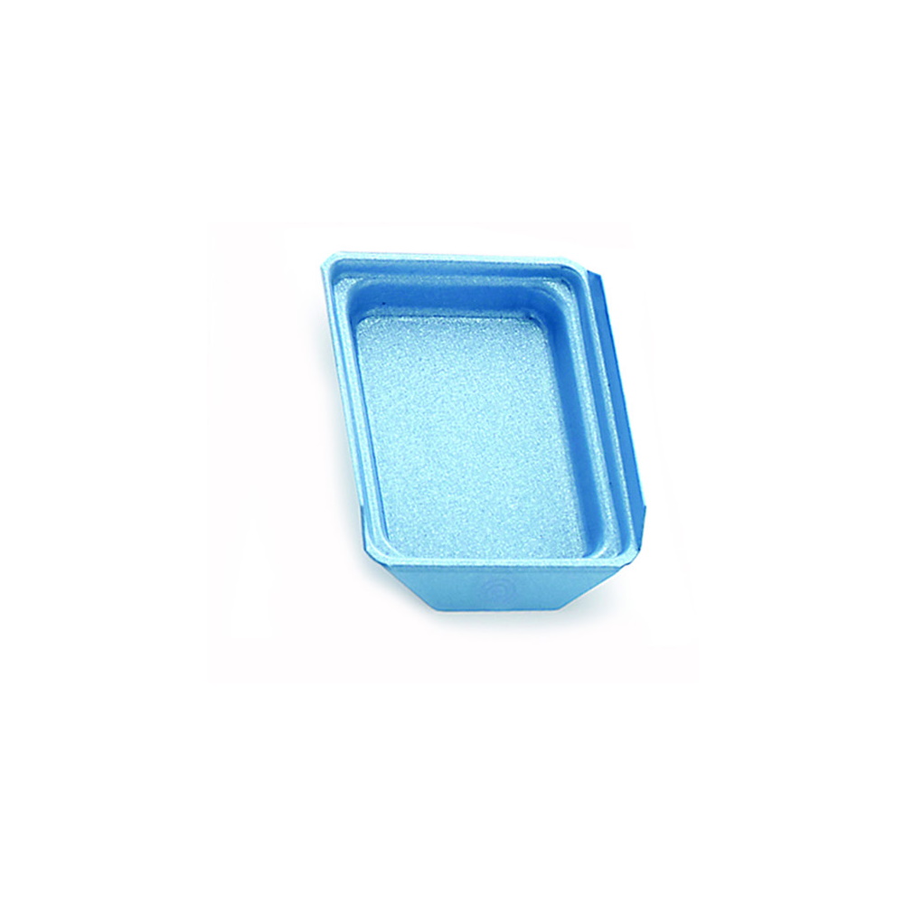
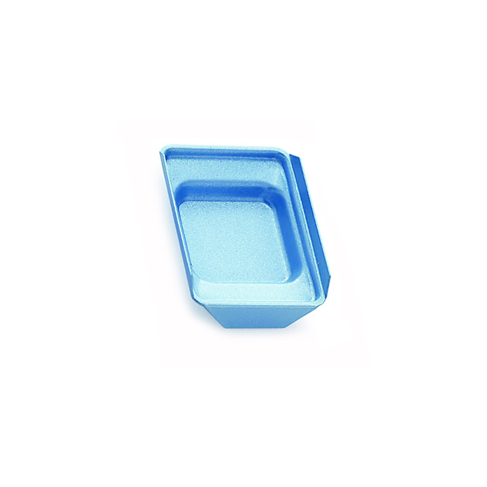
Step up your histology game with the revolutionary Atomic Blue™ base molds! Designed for efficiency and precision, these molds feature a cutting-edge non-stick, nano-layer coating that ensures lightning-fast cooling and effortless block release.
Why Atomic Blue™?
-
-
- Non-Stick Pearlex Coating: Unleash unmatched performance with seamless block release.
- 50% Faster Cooling: Stay ahead with quicker embedding times.
-
Reusable and cost-effective, our molds offer a sustainable solution for consistent, top-tier embedding. Choose from 5mm or 10mm depths to accommodate various specimen sizes, providing versatility without compromise.
Experience hassle-free embedding like never before. Say goodbye to stubborn blocks and elevate your workflow with Atomic Blue™ base molds today!
Enhance your histology embedding process with our 308 stainless steel base molds, engineered for durability, precision, and repeated use. Designed specifically for embedding, these molds ensure optimal specimen orientation for superior sectioning results.
Optimized Design – Enhances uniform embedding and sectioning precision.
Premium 308 Stainless Steel – Resistant to warping & corrosion.
Reusable & Cost-Effective – A long-term solution for consistent, high-quality embedding.
Versatile Depth Options – 5mm or 10mm depths to accommodate various specimen sizes.
Built for the demands of a busy histology lab, these molds provide reliable performance with every use. Upgrade your embedding workflow today!
– Without Preservative
– Not buffered
– pH range 6.5 – 7.0
![]()
STAIN SOLUTION:
| 500 ml | 1 Liter | 1 Gallon | |
| Eosin Y- Phloxine B Working Solution | Part 1083A | Part 1083B | Part 1083C |
Additionally Needed For H&E Staining:
| Hematoxylin and Eosin (H&E) Control Slides | Part 4278 |
| Xylene, ACS | Part 1445 |
| Alcohol, Ethyl Denatured, 100% | Part 10841 |
| Alcohol, Ethyl Denatured, 95% | Part 10842 |
| Hematoxylin Stain, Harris Modified OR Hematoxylin Stain, Harris |
Part 1201 OR Part 12013 |
| Acid Alcohol 1% | Part 10011 |
| Lithium Carbonate, Saturated Aqueous OR Scott Tap Water Substitute |
Part 12215 OR Part 1380 |
| Alcohol, Ethyl Denatured, 70% | Part 10844 |
For storage requirements and expiration date refer to individual product labels.
APPLICATION:
Newcomer Supply Eosin Y-Phloxine B Working Solution provides an enhanced counterstain to hematoxylin and eosin stains, which can be used in either manual or automated staining platforms. With the addition of the phloxine B component, differentiation of muscle, connective tissue and epithelial elements tend to be sharper and better demonstrated than with the traditional eosin Y solution.
Hematoxylin and eosin (H&E) staining is used for screening specimens in anatomic pathology, for research, smears, touch preps and other applications. Its two primary coloring agents stain all cellular material: nuclei (blue), and cytoplasmic elements (pink-red). Popularity of this stain is due to its simplicity, ability to clearly demonstrate a variety of tissue components, dependability, repeatability, and speed of use.
Quality Control: Since hematoxylin and eosin staining is the foundation of the diagnostic process, maintaining quality is of critical importance. Procedures will vary between laboratories depending upon volume of slides, automation vs manual staining, chemical hygiene and solution integrity. The longevity of eosin depends upon these factors and stain quality should be regularly screened with an H&E control slide.
METHOD:
Fixation: Formalin 10%, Phosphate Buffered (Part 1090)
Technique: Paraffin sections cut at 4 microns
Solutions: All solutions are manufactured by Newcomer Supply, Inc.
H&E STAINING PROCEDURE WITH EOSIN Y- PHLOXINE B:
-
- Deparaffinize sections thoroughly in three changes of xylene, 3 minutes each. Hydrate through two changes each of 100% and 95% ethyl alcohols, 10 dips each. Wash well with distilled water.
-
- See Procedure Notes #1 and #2.
-
- Stain with Hematoxylin Stain, Harris Modified (Part 1201) or Hematoxylin Stain, Harris (Part 12013) 1-5 minutes, depending on preference of nuclear stain intensity.
- Wash well in three changes of tap water.
- Differentiate quickly in Acid Alcohol 1%.
-
- Nuclei should be distinct and background very light to colorless.
-
- Rinse well in three changes of tap water.
- Blue slides in Lithium Carbonate, Saturated Aqueous (Part 12215) or Scott Tap Water Substitute (Part 1380) for 60-90 seconds.
- Wash in three changes of tap water; rinse in distilled water.
- Drain excess water; proceed to 70% ethyl alcohol for 10 dips.
- Counterstain in Eosin Y-Phloxine B Working Solution for 1 minute.
- Dehydrate in two changes of 95% ethyl alcohol for 1 minute each and two changes of 100% ethyl alcohol, 10 dips each. Clear in three changes of xylene, 10 dips each; coverslip with compatible mounting medium.
- Deparaffinize sections thoroughly in three changes of xylene, 3 minutes each. Hydrate through two changes each of 100% and 95% ethyl alcohols, 10 dips each. Wash well with distilled water.
RESULTS:
| Nuclei | Blue |
| Erythrocytes and eosinophilic granules | Bright pink to red |
| Cytoplasm and other tissue elements | Various shades of pink |
PROCEDURE NOTES:
-
- Drain slides after each step to prevent solution carry over.
- Do not allow sections to dry out at any point during procedure.
- If using a xylene substitute, follow manufacturer recommendations for deparaffinization and clearing steps.
REFERENCES:
-
- Carson, Freida L. and Christa Cappellano. Histotechnology: A Self-Instructional Text. 5th ed. Chicago: ASCP Press, 2020. 119-120.
- Luna, Lee G. Histopathologic Methods and Color Atlas of Special Stains and Tissue Artifacts. Gaitheresburg, MD: American Histolabs, 1992. 86-87, 91-92.
- Sheehan, Dezna C. and Barbara B. Hrapchak. Theory and Practice of Histotechnology. 2nd ed. St. Louis: Mosby, 1980. 143-144, 153-154.
- Suvarna, S. Kim, Christopher Layton and John D. Bancroft. Bancroft’s Theory and Practice of Histological Techniques. 8th ed. Oxford: Elsevier, 2019. 126-130.
- Modifications developed by Newcomer Supply Laboratory.
The BarrierPaste Brush Applicator PAP Pen is an extremely hydrophobic blocker that is applied via a brush. The barrier forms extra wide lines but dries rapidly.
-
- Suitable for thick cryosections
- 10ml glass bottle with a stirrer magnet and brush
- Shelf life of 18 months
- Each bottle is good for roughly 450 slides
- Available in 6 colors and clear
![]()
SOLUTION:
| 4 X 1 Gallon | |
| Clear Choice | Part 1443C |
Additionally Needed:
| Alcohol, Ethyl Denatured, 70% | Part 10844 |
| Alcohol, Ethyl Denatured, 95% | Part 10842 |
| Alcohol, Ethyl Denatured, 100% | Part 10841 |
| Choice Mounting Medium | Part 1032 |
For storage requirements and expiration date refer to individual bottle labels.
APPLICATION:
Newcomer Supply Clear Choice Xylene Substitute is classified as an aliphatic hydrocarbon that provides a safe alternative to xylene, reduces risks and improves personnel safety in the laboratory. Benefits of Clear Choice Xylene Substitute include:
-
- Odorless, low toxicity, non-irritating and fast drying.
- Gentle on tissue, no brittleness, shrinkage or adverse morphologic changes.
- Compatible with IHC staining.
- Flash point of 66°C/150.8°F (29°C/84°F flash point of xylene)
- Compatible on tissue processors and staining systems.
METHOD:
Fixation: Formalin 10%, Phosphate Buffered (Part 1090)
Technique: Paraffin, frozen sections, smears
PROCESSING PROCEDURE:
-
- Use three Clear Choice Xylene Substitute clearing stations for 60 minutes each.
-
- For two clearing stations, allow 90 minutes per station.
-
- Rotate, filter and/or replace Clear Choice Xylene Substitute stations daily or after processing approximately 1000 blocks.
- Test and optimize Clear Choice Xylene Substitute as a clearing agent in tissue processing schedules prior to standard use.
- Use three Clear Choice Xylene Substitute clearing stations for 60 minutes each.
STAINING PROCEDURE:
-
- Deparaffinize thoroughly in three changes of Clear Choice Xylene Substitute, 3 minutes each. Hydrate through two changes each of 100% and 95% ethyl alcohols, 10 dips each. Wash well with distilled water.
-
- See Procedure Notes #1 and #2.
-
- Proceed with staining protocol.
- Dehydrate in two changes of 95% and three changes of 100% ethyl alcohol. Clear in four changes of Clear Choice Xylene Substitute, 1-2 minutes each change.
- Coverslip with Choice Mounting Medium (Part 1032).
-
- See Procedure Note #3.
-
- Test and optimize Clear Choice Xylene Substitute in staining procedures and automated staining systems prior to standard use.
- Deparaffinize thoroughly in three changes of Clear Choice Xylene Substitute, 3 minutes each. Hydrate through two changes each of 100% and 95% ethyl alcohols, 10 dips each. Wash well with distilled water.
PROCEDURE NOTES:
-
- Deparaffinization and clearing steps may require longer timings than xylene.
- Clear Choice Xylene Substitute may require more frequent changes compared to xylene.
- Test Clear Choice Xylene Substitute compatibility with other mounting mediums prior to use.
-
- If mounting medium displays separation or is not readily miscible, it is incompatible with Clear Choice Xylene Substitute.
-
- Clear Choice Xylene Substitute is not recommended for automated tape coverslippers.
- Clear Choice Xylene Substitute will not remove adhered coverslips as well as xylene.
- Refer to manufacturer’s specifications concerning the use of xylene substitutes on all instrumentation.
REFERENCES:
-
- Dapson, Janet Crookham and Richard W. Dapson. Hazardous Materials in the Histopathology Laboratory: Regulations, Risks, Handling and Disposal. 4th Battle Creek, MI: Anatech, 2005. 150-155, 235.
- Wynnchuk, Maria. “Evaluation of Xylene Substitutes for Paraffin Tissue Processing.” The Journal of Histotechnology 17.2 (1994): 143-149.
- Modifications developed by Newcomer Supply Laboratory.
CaviWipes HP provides a fast 1-minute universal contact time from an alcohol-free hydrogen peroxide formulation that fully qualifies for the EPA’s rigorous Emerging Viral Pathogen Claim. It is ideal for alcohol-sensitive equipment and fast turnarounds.
CAVIWIPES HP SURFACE DISINFECTION TOWELETTES FEATURES AND BENEFITS:
-
- Alcohol-free hydrogen peroxide wipe
- Universal 1-minute contact time against all listed pathogens
- Green lid opens and closes easily and securely and helps dispense wipes without waste
- 1 step cleaner and disinfectant
- Provides disinfections against 67 listed pathogens including MDRO’s
QUALIFICATIONS:
CAVIWIPES HP SURFACE DISINFECTION TOWELETTES LIST OF USES:
-
- Ambulance equipment surfaces
- Animal care facilities
- Bathrooms
- Correctional facilities
- Daycare centers
- Dental offices
- Emergency medical settings
- Emergency vehicles
- Exterior surfaces of anesthesia machines and respiratory therapy equipment
- Health club facilities
- Hospitals
- Infant/child care equipment surfaces
- Interior and exterior surfaces of infant incubators, bassinets
- Isolation areas
- Laboratories
- Laundry rooms
- Neonatal units
- Nursing homes
- Operating rooms
- Ophthalmic and optometric facilities
- Outpatient surgical centers
- Oxygen hoods
- Schools
- Surgical centers
CAVIWIPES HP SURFACE DISINFECTION TOWELETTE KILL CLAIMS:
Mycobacterium
-
- Mycobacterium tuberculosis var: bovis (BCG)
Viruses (Non-Enveloped)
-
- Human Norovirus
- Human Rotavirus
- Rhinovirus Type 37
- Rhinovirus Type 1A
- Human Adenovirus Type 5
- Echovirus Type 11
- Enterovirus Type 71
Viruses (Enveloped)
-
- Herpes simplex virus Type 1 & Type 2
- Avian Influenza A virus (H7N9)
- Avian Influenza A virus (H5N1)
- Influenza A virus (H3N2)
- Influenza B virus (H1N1)
- Parainfluenza virus Type 3
- Hepatitis B virus (HBV)
- Human Hepatitis C virus (HCV)
- Human lmmunodeficiency virus Type 1 (HIV-1)
- Severe Acute Respiratory Syndrome-related Coronavirus 2 (SARS-CoV-2)
- Vaccinia virus
- SARS-Associated Coronavirus (SARs-CoV)
- Cytomegalovirus (CMV)
- Influenza A virus (H1N1) Strains A/4108/2009 and A/04/09
- Human Coronavirus
- Respiratory Syncytial Virus (RSV)
- Measles virus
Pathogenic Fungi
-
- Trichophyton interdigitale
- Candida auris
- Candida albicans
Bacteria
-
- Pseudomonas aeruginosa
- Staphylococcus aureus
- Salmonella enterica
- Bordetella pertussis
- Enterococcus faecium
- Klebsiella aerogenes
- Klebsiella pneumoniae
- Escherichia coli
- Escherichia coli O157:H7
- Burkholderia cepacia
- Stenotrophomonas maltophilia
- Streptococcus pyogenes
- Listeria monocytogenes
- Shigella dysenteriae
- Serratia marcescens
- Bordetella bronchiseptica
- Campylobacter jejuni
- Proteus vulgaris
Multi-Drug-Resistant Bacteria (MDRO)
-
- Penicillin-Resistant Streptococcus pneumoniae (PRSP)
- Vancomycin-Resistant Enterococcus faecalis (VRE)
- Methicillin-Resistant Staphylococcus aureus (MRSA)
- Hospital-Acquired and Community Acquired Methicillin-Resistant
Staphylococcus aureus (HA-MRSA and CA-MRSA) - Vancomycin Intermediate Staphylococcus aureus (VISA)
- Vancomycin Resistant Staphylococcus aureus (VRSA)
- Linezolid-Resistant Staphylococcus aureus (LRSA)
- Carbapenem-Resistant Klebsiella pneumoniae
- Multidrug-Resistant Acinetobacter baumannii (MRAB) Strains
ATCC 19606 and ATCC BAA-1605 - NDM-1 Enterobacter cloacae
- NDM-1 Klebsiella pneumoniae
- MBL Pseudomonas aeruginosa
- NMD-1 Escherichia coli
- ESBL Klebsiella pneumoniae
- ESBL Escherichia coli
- Multidrug-Resistant Klebsiella pneumoniae
- Carbapenem-Resistant Escherichia coli
![]()
PRODUCT SPECIFICATIONS:
Cell Line: Two positive staining cores and one negative staining core from HistoCyte Cell Microarray HCL006
Fixation: Formalin 10%, Phosphate Buffered.
Section/Glass: Paraffin sections cut at 4 microns on Superfrost™ Plus slides.
Storage: 2-8°C in a light deprived and humidity controlled environment.
Expiration: Refer to individual product label.
Intended Use: Research Use Only (RUO).
PRODUCT DESCRIPTION:
The enclosed cell line control slides are developed from a process that allows production of compact cell preparations, cultured from human cell lines, that retain their cellular morphology and are tissue-like in composition. These cell lines are standardized and manufactured to provide consistent results from slide to slide. Each slide contains three, 1.5-2mm diameter cell line cores which demonstrate positive and negative expressions for the specific marker.

-
- A = Breast adenocarcinoma (negative p16 expression)
- B = Cervical adenocarcinoma (positive p16 expression)
- C = Epidermoid carcinoma (positive p16 expression)
APPLICATION:
Newcomer Supply p16 Cell Line Control Slides are appropriate for use in immunohistochemistry (IHC) and in situ hybridization (ISH). The slides may be used to check for reagent performance and troubleshooting of HPV ISH and p16 IHC staining. p16 can serve as a surrogate marker for high-risk HPV in cases of cervical, head and neck and a variety of HPV related carcinomas. It is the responsibility of the end user to determine suitability with their reagents and procedures within their laboratory.
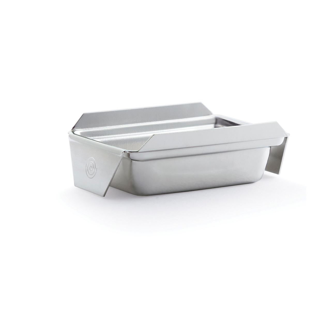
These Histosette II tissue processing & embedding cassettes are suitable for hoppers accepting plastic sleeves such as Thermo Fisher printers. They load in cassette labeling instruments in one simple operation! Molded from acetal, these cassettes keep specimens safely submerged in liquid and are resistant to the chemical action of most histological solvents. The efficient flow-through slots maximize fluid exchange and ensure proper drainage.
PACKAGING OF THE HISTOSETTE II TISSUE PROCESSING & EMBEDDING CASSETTES IN QUICKLOAD SLEEVES:
- 75 cassettes/sleeve; 10 sleeves/case; 750 cassettes/case and 750 covers.
HISTOSETTE II TISSUE PROCESSING & EMBEDDING CASSETTES IN QUICKLOAD SLEEVES:
- Convenient plastic dispensing sleeve compatible with Thermo Fisher printers.
- Efficient flow-through slots maximize fluid exchange and ensure proper drainage.
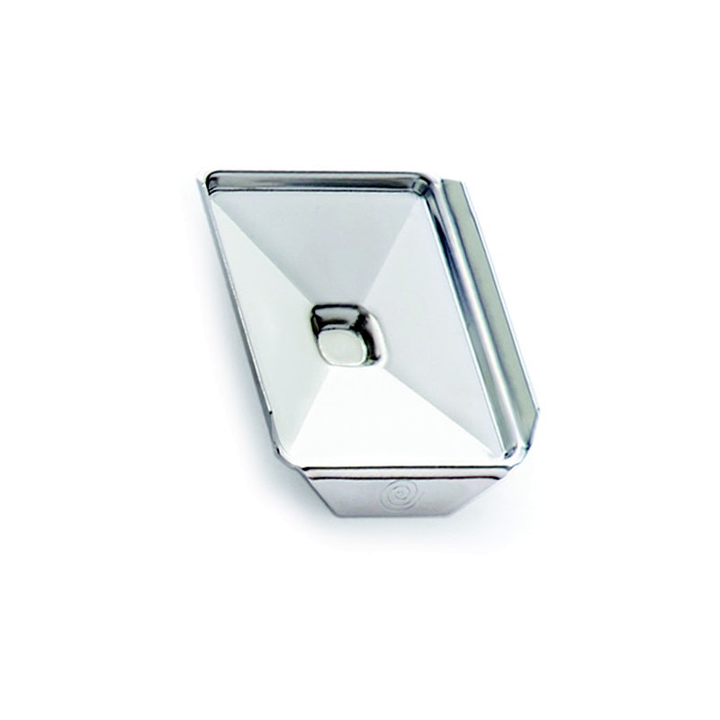
- Lids are packaged separately and have a secure locking device which safely holds the specimen during processing.
- Anterior writing area is at a 45° angle.
- Cassettes also available with QuickLoad Taped Stacks (Part 5131).
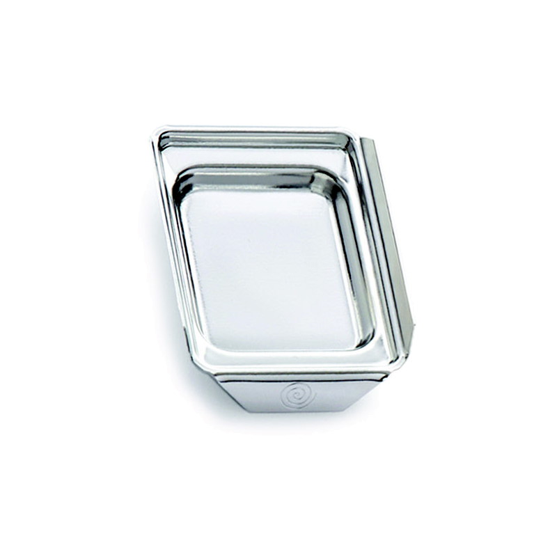
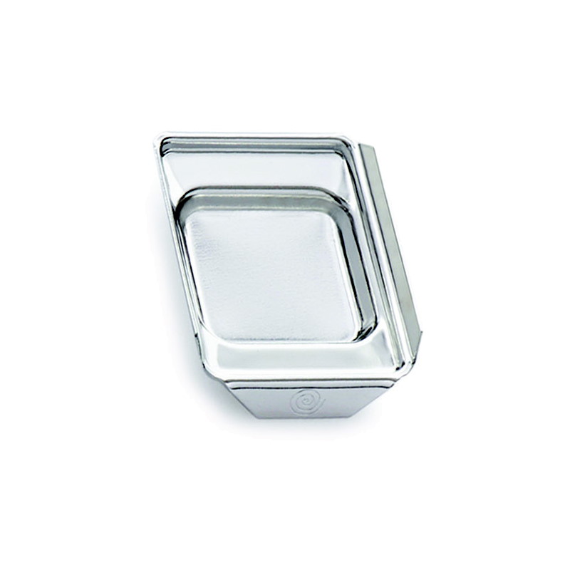
These Histosette II Biopsy tissue processing & embedding cassettes are suitable for hoppers accepting plastic sleeves such as Thermo Fisher printers. They load in cassette labeling instruments in one simple operation! Molded from acetal, these cassettes keep specimens safely submerged in liquid and are resistant to the chemical action of most histological solvents. The efficient flow-through slots maximize fluid exchange and ensure proper drainage.
PACKAGING OF THE HISTOSETTE II BIOPSY TISSUE PROCESSING & EMBEDDING CASSETTES IN QUICKLOAD SLEEVES:
- 75 biopsy cassettes/sleeve; 10 sleeves/case; 750 biopsy cassettes/case and 750 covers.
HISTOSETTE II BIOPSY TISSUE PROCESSING & EMBEDDING CASSETTES IN QUICKLOAD SLEEVES:
- Convenient plastic dispensing sleeve compatible with Thermo Fisher printers.
- 1 mm square openings to maximize fluid exchange.
- Lids are packaged separately and have a secure locking device that safely holds the specimen during processing.
- Anterior writing area is at a 45° angle.
- Cassettes also available with QuickLoad Taped Stacks (Part 5132).
FEATURES OF THE TISSUE FLOTATION BATH – DIGITAL:
-
- Extra deep basin helps lift sections out of the bath easily
- Glass basin is lighted for easy tissue placement
- Large slide placement area
- Quiet fan-less design
- High Definition touch screen
- Temperature accuracy within 1°C
- Nonvolatile memory retains the last temperature set point after turning off
- Timer and alarm reminder
- Auto turn-on/turn-off function
SPECIFICATIONS:
-
- Temperature Range: 0°C-70°C
- Voltage: AC120V ±10%, 60Hz±1Hz
- Power: 200 Watts
- Dimensions: 12 ¾”L x 13 ¼”W x 6 ½”H
- Glass Basin Dimensions: 8 ¼”L x 6 ½”W x 2 ¼”H
- Relative Humidity: < 80%
- Temperature Accuracy: ±1°C
- Heating Time: 20 minutes to heat up to 54°C
- Exterior Material: Epoxy Painted Cold Rolled Steel
- Large slide placement area: Accommodates 8 slides
WARRANTY DETAILS:
-
- Two year warranty
Manual for Tissue Flotation Bath TFB200101

HistoCyte Laboratories’ NTRK Analyte Control contains two cell lines that demonstrate positive and negative expression of NTRK. It specifically expresses wild type (WT) TrkA, that is recognized by the pan-NTRK antibodies as the WT Trk proteins and fusion proteins share a highly homologous c-terminus, within which the tyrosine kinase domain resides. Ideal for use as a same slide quality control in immunohistochemistry (IHC) to demonstrate the reagents have been applied to the slide. These cell lines are derived from the following tumors:
Cell line A: Breast adenocarcinoma
Cell line B: Large cell lymphoma
- Cells are fixed in 10% neutral buffered formalin and paraffin wax embedded.
- Sections are cut at 4µm, mounted on positively charged slides and dried overnight at 37ºC.
- Cell microarrays (CMA) contain cores that are 1.5-2mm in diameter and 3-3.5mm in length. It is possible to obtain over 300 sections depending on thickness.
- Not suitable for FISH as there is no translocation in the cell line. It is WT NTRK1.
HistoCyte Laboratories’ MSH6 Analyte Control contains 2 cell lines, one with intact expression for MSH6 and one with loss of expression for MSH6. This two core product is a cost effective means of controlling for MSH6 assays over the four core MMR Analyte Control. However, it cannot be used with other MMR assays. These cell lines are derived from the following tumors:
Cell line A: Breast adenocarcinoma
Cell line B: Colon adenocarcinoma
- Cells are fixed in 10% neutral buffered formalin and paraffin wax embedded.
- Sections are cut at 4µm, mounted on positively charged slides and dried overnight at 37ºC.
- Cell microarrays (CMA) contain cores that are 1.5-2mm in diameter and 3-3.5mm in length. It is possible to obtain over 300 sections depending on thickness.
HistoCyte Laboratories’ MSH2 Analyte Control contains 2 cell lines, one with intact expression for MSH2 and one with loss of expression for MSH2. This two core product is a cost effective means of controlling for MSH2 assays over the four core MMR Analyte Control. However, it cannot be used with other MMR assays. These cell lines are derived from the following tumors:
Cell line A: Breast adenocarcinoma
Cell line B: Colon adenocarcinoma
- Cells are fixed in 10% neutral buffered formalin and paraffin wax embedded.
- Sections are cut at 4µm, mounted on positively charged slides and dried overnight at 37ºC.
- Cell microarrays (CMA) contain cores that are 1.5-2mm in diameter and 3-3.5mm in length. It is possible to obtain over 300 sections depending on thickness.
HistoCyte Laboratories’ MMR Analyte Control consists of four different cell lines with loss of and intact expression for MLH1, PMS2, MSH2 and MSH6. This product is a convenient quality control solution being suitable for use with all the MMR assays, regardless of which assay is used. These cell lines are derived from the following tumors:
Cell line A: Breast adenocarcinoma
Cell line B: Prostate carcinoma
Cell line C: Colon adenocarcinoma
Cell line D: Colon adenocarcinoma
- Cells are fixed in 10% neutral buffered formalin and paraffin wax embedded.
- Sections are cut at 4µm, mounted on positively charged slides and dried overnight at 37ºC.
- Cell microarrays (CMA) contain cores that are 1.5-2mm in diameter and 3-3.5mm in length. It is possible to obtain over 300 sections depending on thickness.
HistoCyte Laboratories’ MLH1/PMS2 Analyte Control contains 2 cell lines, one with intact expression for MLH1 and PMS2 and one with loss of expression for MLH1 and PMS2. This two core product is a cost effective means of controlling for MLH1 and PMS2 assays over the four core MMR Analyte Control. However, it cannot be used with the other MMR assays. These cell lines are derived from the following tumors:
Cell line A: Breast adenocarcinoma
Cell line B: Prostate carcinoma
- Cells are fixed in 10% neutral buffered formalin and paraffin wax embedded.
- Sections are cut at 4µm, mounted on positively charged slides and dried overnight at 37ºC.
- Cell microarrays (CMA) contain cores that are 1.5-2mm in diameter and 3-3.5mm in length. It is possible to obtain over 300 sections depending on thickness.
HistoCyte Laboratories’ HPV/p16 Analyte Control contains 3 cell lines that demonstrate high, medium and negative expression of high-risk human papillomavirus types 16 and 18 by in situ hybridization (ISH). The same cell lines also demonstrate high, intermediate (heterogeneous) and negative expression of p16 by immunohistochemistry (IHC). Ideal for use as a same slide control for HPV ISH and p16 IHC to demonstrate assay efficacy. These cell lines are derived from the following tumors:
Cell line A: Human breast adenocarcinoma
Cell line B: Human cervical adenocarcinoma
Cell line C: Human epidermoid carcinoma
- Cells are fixed in 10% neutral buffered formalin and paraffin wax embedded.
- Sections are cut at 4µm, mounted on positively charged slides and dried overnight at 37ºC.
- Cell microarrays (CMA) contain cores that are 1.5-2mm in diameter and 3-3.5mm in length. It is possible to obtain over 300 sections depending on thickness.
![]()
HistoCyte Laboratories’ multi-purpose Breast Analyte Control is a qualitative control that contains two cell lines that demonstrate positive and negative expression of ER, PR and HER2. Ideal for use as a same slide control in immunohistochemistry (IHC) to demonstrate the reagents have been correctly applied to the slide. These cell lines are derived from the following tumors:
Cell line A: Osteosarcoma
Cell line B: Breast ductal carcinoma
- Cells are fixed in 10% neutral buffered formalin and paraffin wax embedded.
- Sections are cut at 4µm, mounted on positively charged slides and dried overnight at 37ºC.
- Cell microarrays (CMA) contain cores that are 1.5-2mm in diameter and 3-3.5mm in length. It is possible to obtain over 300 sections depending on thickness.
HistoCyte Laboratories’ BRAF V600E Analyte Control consists of two cores negative and positive for expression of BRAF V600E. This provides a cost effective means on controlling for BRAF V600E in immunohistochemistry. These cell lines are derived from the following tumors:
Cell line A: Breast ductal carcinoma
Cell line B: Malignant melanoma
- Cells are fixed in 10% neutral buffered formalin and paraffin wax embedded.
- Sections are cut at 4µm, mounted on positively charged slides and dried overnight at 37ºC.
- Cell microarrays (CMA) contain cores that are 1.5-2mm in diameter and 3-3.5mm in length. It is possible to obtain over 300 sections depending on thickness.
HistoCyte Laboratories’ ALK-Lymphoma Analyte Control contains two cell lines that demonstrate positive and negative expression of NPM-ALK associated lymphoma. This is a cost effective same slide quality control for use in immunohistochemistry (IHC) to demonstrate the reagents have been applied to the slide. These cell lines are derived from the following tumors:
Cell line A: Breast Adenocarcinoma
Cell line B: Anaplastic large cell lymphoma
- Cells are fixed in 10% neutral buffered formalin and paraffin wax embedded.
- Sections are cut at 4µm, mounted on positively charged slides and dried overnight at 37ºC.
- Cell microarrays (CMA) contain cores that are 1.5-2mm in diameter and 3-3.5mm in length. It is possible to obtain over 300 sections depending on thickness.
HistoCyte Laboratories’ ROS1 Analyte ControlDR is the dynamic range version of the ROS1 control, containing an additional cell line with very low expression of ROS1. These cell lines are derived from the following tumors:
Cell line A: Breast Adenocarcinoma
Cell line B: Glioblastoma
Cell line C: Lung adenocarcinoma
- Cells are fixed in 10% neutral buffered formalin and paraffin wax embedded.
- Sections are cut at 4µm, mounted on positively charged slides and dried overnight at 37ºC.
- Cell microarrays (CMA) contain cores that are 1.5-2mm in diameter and 3-3.5mm in length. It is possible to obtain over 300 sections depending on thickness.
HistoCyte Laboratories’ PR Dynamic Range Analyte Control contains four cores, offering a range of expression for PR: Negative, Low/Intermediate, Intermediate/High, and High. These cell lines are derived from the following tumors:
Cell line A: Ductal Carcinoma
Cell line B: Breast Adenocarcinoma
Cell line C: Ductal Carcinoma
Cell line D: Ductal Carcinoma
- Cells are fixed in 10% neutral buffered formalin and paraffin wax embedded.
- Sections are cut at 4µm, mounted on positively charged slides and dried overnight at 37ºC.
- Cell microarrays (CMA) contain cores that are 1.5-2mm in diameter and 3-3.5mm in length. It is possible to obtain over 300 sections depending on thickness.
![]()
HistoCyte Laboratories’ PD-L1 Analyte ControlDR consists of four different cell lines with PD-L1 expression levels of high, intermediate, low and negative. Ideal for use as a same slide control for PD-L1 to demonstrate the sensitivity of your assay and improve confidence in the result. These cell lines are derived from the following tumors:
Cell line A: Breast ductal carcinoma
Cell line B: Osteosarcoma
Cell line C: Fibrosarcoma
Cell line D: T-cell non-Hodgkin lymphoma
- Cells are fixed in 10% neutral buffered formalin and paraffin wax embedded.
- Sections are cut at 4µm, mounted on positively charged slides and dried overnight at 37ºC.
- Cell microarrays (CMA) contain cores that are 1.5-2mm in diameter and 3-3.5mm in length. It is possible to obtain over 300 sections depending on thickness.
HistoCyte Laboratories’ HPV/p16 Analyte ControlDR contains 4 cell lines that demonstrate a full Dynamic Range of expression for high risk human papillomavirus types 16 and 18: high, medium, low and negative gene status by ISH. The same cell lines demonstrate homogenous, heterogenous and negative expression of p16. Ideal for use as same slide control for HPV in situ hybridization (ISH) for strains 16 and 18 and in p16 immunohistochemistry (IHC). These cell lines are derived from the following tumors:
Cell line A: Human breast adenocarcinoma
Cell line B: Human cervical squamous cell carcinoma
Cell line C: Human cervical adenocarcinoma
Cell line D: Human epidermoid carcinoma
- Cells are fixed in 10% neutral buffered formalin and paraffin wax embedded.
- Sections are cut at 4µm, mounted on positively charged slides and dried overnight at 37ºC.
- Cell microarrays (CMA) contain cores that are 1.5-2mm in diameter and 3-3.5mm in length. It is possible to obtain over 300 sections depending on thickness.
HistoCyte Laboratories’ ER Dynamic Range Analyte Control contains four cores, offering a full range of expression for ER: Negative, Low, Intermediate, and High. These cell lines are derived from the following tumors:
Cell line A: Osteosarcoma
Cell line B: Urothelial carcinoma
Cell line C: Non-small cell lung carcinoma
Cell line D: Breast adenocarcinoma
- Cells are fixed in 10% neutral buffered formalin and paraffin wax embedded.
- Sections are cut at 4µm, mounted on positively charged slides and dried overnight at 37ºC.
- Cell microarrays (CMA) contain cores that are 1.5-2mm in diameter and 3-3.5mm in length. It is possible to obtain over 300 sections depending on thickness.
HistoCyte Laboratories’ multipurpose Breast Analyte ControlDR contains five cell lines that demonstrate a Dynamic Range of 0, 1+ and 3+ for HER2. For ER and PR a variety of staining provides a degree of sensitivity over a standard control. Ideal for use in a laboratory where one control can be used across multiple assays as a same slide control in immunohistochemistry (IHC). These cell lines are derived from the following tumors:
Cell line A: Osteosarcoma
Cell line B: Breast ductal carcinoma
Cell line C: Breast ductal carcinoma
Cell line D: Breast ductal carcinoma
Cell line E: Breast ductal carcinoma
- Cells are fixed in 10% neutral buffered formalin and paraffin wax embedded.
- Sections are cut at 4µm, mounted on positively charged slides and dried overnight at 37ºC.
- Cell microarrays (CMA) contain cores that are 1.5-2mm in diameter and 3-3.5mm in length. It is possible to obtain over 300 sections depending on thickness.
HistoCyte Laboratories’ ALK-Lung Analyte Control is a qualitative control containing two cell lines that demonstrate positive and negative expression of EML4-ALK. This fusion protein is commonly associated with lung cancer. Ideal for use as a same slide control in immunohistochemistry (IHC) or fluorescence in situ hybridization (FISH) to demonstrate the reagents have been correctly applied to the slide. These cell lines are derived from the following tumors:
Cell line A: Breast Adenocarcinoma
Cell line B: Lung Adenocarcinoma
- Cells are fixed in 10% neutral buffered formalin and paraffin wax embedded.
- Sections are cut at 4µm, mounted on positively charged slides and dried overnight at 37ºC.
- Cell microarrays (CMA) contain cores that are 1.5-2mm in diameter and 3-3.5mm in length. It is possible to obtain over 300 sections depending on thickness.
HistoCyte Laboratories’ ALK Analyte ControlDR consists of four different cell lines expressing WT and ALK fusion proteins. These cell lines are derived from the following tumors:
Cell line A: Breast adenocarcinoma
Cell line B: Glioblastoma
Cell line C: Lung adenocarcinoma
Cell line D: Anaplastic large cell lymphoma
- Cells are fixed in 10% neutral buffered formalin and paraffin wax embedded.
- Sections are cut at 4µm, mounted on positively charged slides and dried overnight at 37ºC.
- Cell microarrays (CMA) contain cores that are 1.5-2mm in diameter and 3-3.5mm in length. It is possible to obtain over 300 sections depending on thickness.
Discover the ultimate paraffin wax trimmer, meticulously crafted to elevate every histologist’s experience. With a spacious working surface, effortlessly tidy and trim numerous FFPE tissue blocks simultaneously.
Its intuitive design simplifies operation, featuring a rotary knob serving as both the power switch and temperature controller. Within 5-10 minutes, it reaches optimal temperature, primed for use.
The innovative hot plate design, complete with ample outer grooves, swiftly channels melted paraffin into the waste bin, magnetically attached for seamless disposal. Transform your workflow with this unparalleled tool—it’s an absolute delight to operate!

FEATURES OF THE PARAFFIN WAX TRIMMER – DIGITAL, FOUR E’S:
-
- LED display and rotary knob for continuous temperature adjustment
- Set temperature is reached in approximately 5 minutes
- Large working surface (11.5″ x 5.25″) allowing multiple cassettes to be trimmed
- Large outer grooved tracks on hot plate for superior wax drainage
- Drip tray is attached magnetically and includes tray liners for easy maintenance
- Power failure recovery mode and over-temperature protection
- Safety notification on display alerts users the surface is hot until plate cools down below 50°C
SPECIFICATIONS:
-
- Temperature Range: 30°C – 99°C
- Temperature accuracy ±1°C
- Voltage: 110V ± 10%; 60Hz
- Power: 250W
- Weight: 3.0 kg (6.6 lb)
- Dimensions: 13 ¾”L x 6 ¼”W x 8 ¼”H
WARRANTY DETAILS:
-
- Two year warranty
Manual for Paraffin Wax Trimmer – Four E’s
The Slide Filing System Metal Cabinets are used in histology laboratories and hospitals and are designed for long term storage of large quantities of microscope slides.


SPECIFICATIONS OF THE SLIDE FILING SYSTEM METAL CABINETS:
-
- Six drawers holding up to 5,000 standard microscope slides per cabinet
- Safe stacking with interlocking system
- Stack up to 12 units high
- Durable cold rolled steel construction with epoxy powder coating
- Each drawer comes with two foam blocks to maintain slides upright when drawers are partially filled
- Base available to raise bottom cabinet
DIMENSIONS OF THE SLIDE FILING SYSTEM METAL CABINETS AND BASE:
-
- Cabinet Dimensions: 18 ¾”L x 15 ¾”W x 5″H
- Base Dimensions: 18 ¾”L x 15 ¾”W x 5″H
CaviWipes 2.0 is a multi-purpose disinfectant for use in cleaning and disinfecting hard, non-porous, inanimate surfaces and non-critical instruments in hospitals, laboratories and other critical care areas where environmental control of cross-contamination between treated surfaces is important.
CAVIWIPES 2.0 SURFACE DISINFECTION TOWELETTES FEATURES & BENEFITS:
- 2-minute universal contact time for bacteria, viruses, and pathogenic fungi (Candida auris)
- Proven effective against SARS-CoV-2 on hard non-porous surfaces
- 1 step disinfecting (if no visible debris present on surface)
- Effective against 60 relevant pathogens including Norovirus and Candida auris for added confidence in preventing healthcare associated infections.
- Excellent Material Compatibility – Compatible with most non-porous, hard surfaces commonly used in clinical settings
QUALIFICATIONS:
- EPA’s Emerging Viral Pathogens claim for all virus types (enveloped, large and small non-enveloped viruses) to meet current and potentially future infection prevention needs
- On EPA List Q
CAVIWIPES 2.0 SURFACE DISINFECTION TOWELETTES LIST OF USES:
- Ambulance equipment surfaces
- Animal care facilities
- Bathrooms
- Correctional facilities
- Daycare centers
- Dental offices
- Emergency medical settings
- Emergency vehicles
- Exterior surfaces of anesthesia machines and respiratory therapy equipment
- Health club facilities
- Hospitals
- Infant/child care equipment surfaces
- Interior and exterior surfaces of infant incubators, bassinets
- Isolation areas
- Laboratories
- Laundry rooms
- Neonatal units
- Nursing homes
- Operating rooms
- Ophthalmic and optometric facilities
- Outpatient surgical centers
- Oxygen hoods
- Schools
- Surgical centers
CAVIWIPES 2.0 SURFACE DISINFECTION TOWELETTES KILL CLAIMS:
2 Minute Efficacy Against
- Adenovirus Type 2
- Echovirus Type 11
- Enterovirus Type 71
- Human Rotavirus
- Norovirus
- Adenovirus Type 5
- Rhinovirus Type 1A
Pathogenic Fungi
- Candida auris
Multi-Drug-Resistant Bacteria
- Penicillin-Resistant Streptococcus pneumoniae (PRSP)
- Vancomycin-Resistant Enterococcus faecalis (VRE)
- Methicillin-Resistant Staphylococcus aureus (MRSA)
- Carbapenem-resistant Klebsiella pneumoniae (CRKP)
- Multidrug-Resistant Acinetobacter baumannii (MRAB)
- NDM-1 Enterobacter cloacae
- Vancomycin-Intermediate Staphylococcus aureus (VISA)
- Methicillin-Resistant Staphylococcus epidermidis
- Multi-Drug Resistant Pseudomonas aeruginosa
- ESBL Klebsiella pneumoniae
- ESBL Escherichia coli
Bacteria
- Acinetobacter baumannii
- Bordetella pertussis
- Burkholderia cepacia
- Enterococcus faecium
- Escherichia coli O157:H7
- Klebsiella pneumoniae
- Legionella pneumophila
- Micrococcus luteus
- Moraxella catarrhalis
- Pseudomonas aeruginosa
- Salmonella enterica
- Staphylococcus aureus
- Stenotrophomonas maltophilia
- Streptococcus mutans
- Streptococcus pyogenes
- Klebsiella aerogenes
- Serratia marcescens
- Campylobacter jejuni
- Listeria monocytogenes
- Shigella dysenteriae
- Chlamydophila pneumoniae
- Flavobacterium columnare
- Mycobacterium tuberculosis (TB)
Enveloped Viruses
- Herpes simplex virus Type 1
- Herpes simplex virus Type 2
- Influenza A virus
- Varicella-zoster virus
- Cytomegalovirus
- SARS-associated Coronavirus
- SARS-CoV-2
- SARS-CoV-2-UK Variant
- SARS-CoV-2 South African Variant
- SARs-CoV-2-California Variant
- SARS-CoV-2 Delta Variant
- Influenza B virus
- Parainfluenza virus Type 3
- Human orthopneumovirus
- Measles virus
- Hepatitis C Virus (HCV)
- Hepatitis B Virus (HBV)
- Human Immunodeficiency Virus Type 1 (HIV-1)
![]()
SOLUTIONS:
| Melanin Pigment Removal | 250 ml | 500 ml | 1 Liter | 4 Liters | 1 Gallon |
| Potassium Permanganate 0.25%, Aqueous | Part 133931A | Part 133931B | |||
| Oxalic Acid 5%, Aqueous | Part 1293A | Part 1293B | |||
| Mercury Pigment Removal | |||||
| Melanin Control Slides | Part 4430 | ||||
| Iodine, Gram, Aqueous OR Iodine, Lugol’s, Aqueous |
Part 1140A OR Part 12092A |
Part 1140C OR Part 12092B |
Part 1140E
|
||
| Sodium Thiosulfate 5%, Aqueous | Part 1389A | Part 1389B | |||
| Formalin Pigment Removal | |||||
| Picric Acid, Saturated Alcoholic | Part 1337A | Part 1337B | Part 1337C |
For storage requirements and expiration date refer to individual product labels.
APPLICATIONS:
Newcomer Supply Pigment and Artifact Pigment Removal Technical Memo provides procedures for removal of pigments, both naturally occurring and artifact from tissue sections.
Melanin pigment: naturally occurs and is produced by melanocytes that provides skin, hair and eyes with color. When melanin pigment obscures cellular detail, it can be bleached with potassium permanganate and oxalic acid solutions.
Artifact pigments: are produced in tissues during processing, often a result of fixation. Microscopically, these pigments usually appear to lie on top of the tissue and not within the cell.
- Mercury pigment: is deposited after exposure to fixatives containing mercuric chloride. Sections must be treated for mercury pigment removal prior to staining.
- Formalin pigment: results when acidic formalin solutions react with blood rich tissues such as spleen and areas of hemorrhage, forming brown or brownish-black crystalline birefringent substances. The use of Formalin 10%, Phosphate Buffered (Part 1090) assists in minimizing formalin pigment deposition.
METHOD:
Technique: Paraffin sections on adhesive slides
-
-
- See Procedure Note #1.
-
Solutions: All solutions are manufactured by Newcomer Supply, Inc.
PROCEDURES:
-
- Deparaffinize sections thoroughly in three changes of xylene, 3 minutes each. Hydrate through two changes each of 100% and 95% ethyl alcohols, 10 dips each. Wash well with distilled water.
- Melanin Pigment Removal:
-
- Two Melanin Control Slides (Part 4430) and two patient slides are needed.
- Label one control slide and one patient slide “with”.
- Label the other control slide and patient slide “without”. Set aside for Step h.
- Bleach “with” sections in Potassium Permanganate 0.25%, Aqueous (Part 133931) for 5 to 20 minutes.
- Rinse in several changes of distilled water.
- Clear in Oxalic Acid 5%, Aqueous (Part 1293) for 1-2 minutes or until sections turn white.
- Wash in gently running tap water for 10 minutes.
- Stain as desired; including untreated melanin control and untreated patient slides labeled “without”.
- See Procedure Note #2.
-
-
- Mercury Pigment Removal:
-
- Treat sections with Iodine, Gram, Aqueous (Part 1140) or Iodine, Lugol’s, Aqueous (Part 12092) for 10 minutes.
- Wash briefly in running tap water.
- Place in Sodium Thiosulfate 5%, Aqueous (Part 1389) for 3 minutes.
- Wash in gently running tap water for 10 minutes.
- Stain as desired.
-
- Formalin Pigment Removal:
-
- Treat sections with Picric Acid, Saturated Alcoholic (Part 1337) for 10 minutes to 3 hours.
- Wash in gently running tap water for 10 minutes.
- Stain as desired.
- See Procedure Note #3.
-
- Mercury Pigment Removal:
PROCEDURE NOTES:
-
- Pigment removal procedures are harsh on tissues sections. The use of adhesive slides (Part 5070, 5079 or 6203) is recommended to ensure tissue adherence.
- The darker the melanin pigment the longer bleach will take to decolorize the pigment.
- Timing to remove formalin pigment will vary, depending on amount of pigment present in sections.
REFERENCES:
-
- Bancroft, John D. and Marilyn Gamble. Theory and Practice of Histological Techniques. 6th ed. Oxford: Churchill Livingstone Elsevier, 2008. 252-253.
- Carson, Freida L. and Christa Hladik. Histotechnology: A Self-Instructional Text. 3rd ed. Chicago, Ill.: American Society of Clinical Pathologists, 2009. 23-24, 254-255.
- Sheehan, Dezna C. and Barbara B. Hrapchak. Theory and Practice of Histotechnology. 2nd ed. St. Louis: Mosby, 1980. 130, 214, 220-221.
- Modifications developed by Newcomer Supply Laboratory.
HistoCyte Laboratories’ ROS1 Analyte Control consists of two cores negative and positive for expression of ROS1. This provides a cost effective means on controlling for ROS1 in immunohistochemistry and fluorescence in situ hybridization. These cell lines are derived from the following tumors:
Cell line A: Breast adenocarcinoma
Cell line B: Lung adenocarcinoma
- Cells are fixed in 10% neutral buffered formalin and paraffin wax embedded.
- Sections are cut at 4µm, mounted on positively charged slides and dried overnight at 37ºC.
- Cell microarrays (CMA) contain cores that are 1.5-2mm in diameter and 3-3.5mm in length. It is possible to obtain over 300 sections depending on thickness.
![]()
HistoCyte Laboratories’ specialized HER2 control has a full dynamic range of expression. Enhanced from multi-purpose breast analyte control to now include a 2+ cell line, this is specifically aimed at those wanting the most reliable, sensitive and accurate control for HER2. These cell lines are derived from the following tumors:
Cell line A: Breast adenocarcinoma
Cell line B: Breast adenocarcinoma
Cell line C: Gastric adenocarcinoma
Cell line D: Breast adenocarcinoma
- Cells are fixed in 10% neutral buffered formalin and paraffin wax embedded.
- Sections are cut at 4µm, mounted on positively charged slides and dried overnight at 37ºC.
- Cell microarrays (CMA) contain cores that are 1.5-2mm in diameter and 3-3.5mm in length. It is possible to obtain over 300 sections depending on thickness.
The Slide Staining Tray, 18 Place is ideal for clinical and research laboratory staining. It holds 18 standard microscope slides (76x26mm) securely in place for staining, rinsing and dry application.
FEATURES OF THE SLIDE STAINING TRAY, 18 PLACE:
-
- Durably constructed from solvent-resistant polypropylene blend
- Non-skid silicone feet keep tray in place
- Two ways to empty stain liquid: pour spouts on each corner or use drain hole
- Drain hole included with plug
- Equipped with handle cut-outs for easy handling
- Black lid included for light sensitive applications
- Humidity retention wells for specialized staining needs
- Proudly made in the USA
SPECIFICATIONS:
-
- Color: Black
- Capacity: 18 standard microscope slides (76 x 26 mm)
- Tray material: polypropylene with talc
- Feet & plug material: Silicone
- Operating temperature: 0 to 40°C
- Storage temperature: -20 to 60°C
- Tray Dimensions: 16.1 x 8.9 x 1.6 in (41 x 22.5 x 4 cm)
- Cover Dimensions: 15.7 x 8.4 x 0.4 in (39.8 x 21.4 x 1.1 cm)
INCLUDES:
-
- 1 Black Base/Tray
- 1 Black Cover
- 2 Drain Plugs
The Graduated Beakers are an excellent choice for mixing, pouring, stirring and measuring solutions in many laboratory operations.
SPECIFICATIONS OF THE GRADUATED BEAKERS:
-
-
- Made of translucent polypropylene for general laboratory work
- Bold, printed, dual scale graduations
- Single non-drip spout
- Chemically resistant to most acids, bases and many common solvents
- Autoclavable at 121°C
- Beakers without handles stack to save storage space
-
The Graduated Cylinders offer precise fluid measurements in many laboratory operations.
SPECIFICATIONS OF THE GRADUATED CYLINDERS:
-
- Made of translucent polypropylene for general laboratory work
- Easy to read molded graduations
- Wide openings with tapered pour spouts and a hexagonal base
- Chemically resistant to most acids, bases and many common solvents
- Working temperature range: 4° to 60°C
![]()
PRODUCT SPECIFICATIONS:
Tissue: Positive staining animal organ.
Fixation: Formalin 10%, Phosphate Buffered (Part 1090).
Section/Glass: Paraffin sections cut at 4 microns on Superfrost™ Plus slides.
Quality Control Stain: Grocott Methenamine Silver quality control stained slide(s) included.
Reactivity: Guaranteed product specific reactivity for one year from date of receipt. Revalidate after one year to verify continued reactivity.
Storage: 15-30°C in a light deprived and humidity controlled environment.
Intended Use: To verify histological techniques and reagent reactivity.
Before using unstained control slides, review the enclosed stained slide(s) to ensure that this tissue source is acceptable for testing needs.
CONTROL SLIDE VALIDATION:
| With Fungus, Grocott Methenamine Silver (GMS) Stain Kit: | Part 9121A/B | Individual Stain Solution | |
| Solution A: | Chromic Acid 5%, Aqueous | 250/500 ml | Part 10341 |
| Solution B: | Sodium Bisulfite 1%, Aqueous | 250/500 ml | Part 13821 |
| Solution C: | Silver Nitrate | 125/250 ml | Part 1142 |
| Solution D: | Methenamine Borate | 125/250 ml | Part 1142 |
| Solution E: | Gold Chloride 0.1%, Aqueous | 250/500 ml | Part 11285 |
| Solution F: | Sodium Thiosulfate 2%, Aqueous | 250/500 ml | Part 13888 |
| Solution G: | Light Green SF Yellowish Stain 0.02%, Aqueous | 250/500 ml | Part 12204 |
APPLICATION:
Newcomer Supply Fungus, GMS, Aspergillus, Animal Control Slides are for the positive histochemical staining of fungal organisms in tissue sections. The organism morphology is consistent with Aspergillus sp.
PRESTAINING PREPARATION:
-
- Heat dry sections in oven according to your laboratory protocol.
- All glassware/plasticware must be acid cleaned prior to use.
-
- See Procedure Notes #1 and #2.
-
- Prepare Silver-Methenamine Working Solution and mix well:
-
- Solution C: Silver Nitrate 20 ml
- Solution D: Methenamine Borate 20 ml
-
- Preheat Silver-Methenamine Working Solution to 45°-60°C in a water bath 20-30 minutes before use.
-
- Maintain solution between 45°-60°C to minimize precipitate.
- Do not preheat if using Microwave Modification; Step 11.
-
NEWCOMER SUPPLY VALIDATION PROCEDURE:
-
- Deparaffinize sections thoroughly in three changes of xylene, 3 minutes each. Hydrate through two changes each of 100% and 95% ethyl alcohols, 10 dips each. Wash well with distilled water.
-
- See Procedure Notes #3 and #4.
-
- Oxidize in Solution A: Chromic Acid 5%, Aqueous for 1 hour.
- Deparaffinize sections thoroughly in three changes of xylene, 3 minutes each. Hydrate through two changes each of 100% and 95% ethyl alcohols, 10 dips each. Wash well with distilled water.
Microwave Modification: See Procedure Note #5.
-
-
-
- Microwave Solution A: Chromic Acid 5%, Aqueous without slides in a plastic Coplin jar (Part 5184) for 1 minute at 60°C. Add slides to heated Solution A and oxidize for 10 minutes.
-
-
-
- Wash well in running tap water; rinse in distilled water.
- Place in Solution B: Sodium Bisulfite 1%, Aqueous for 1 minute.
- Wash for 5 minutes in running tap water; rinse well in distilled water.
- Incubate slides in preheated Silver-Methenamine Working Solution (Step #4) at 45°-60°C or at room temperature, for 12-18 minutes until sections appear paper-bag brown.
-
- Periodically remove control, rinse in warm distilled water, check microscopically for adequate silver impregnation. Fungi should be dark brown.
- If organisms are not sufficiently dark, return slides to warm silver solution. Recheck at 2-3 minute intervals until desired intensity is achieved.
- Staining at room temperature will require longer incubation.
-
-
- Microwave Modification:
-
- Incubate slides in a plastic Coplin jar containing Silver-Methenamine Working Solution. Microwave for 5 minutes at 45°C.
- Check microscopically for adequate development.
- If additional incubation is required, return slides to warm silver solution. Recheck at 3-5 minute intervals.
-
- Rinse in three to four changes of distilled water.
-
- Never use tap water at this step.
-
- Tone in Solution E: Gold Chloride 0.1%, Aqueous until sections turn gray; 10-30 seconds.
- Rinse well in distilled water.
- Remove unreduced silver in Solution F: Sodium Thiosulfate 2%, Aqueous for 2 minutes.
- Wash in running tap water for 5 minutes; rinse in distilled water.
- Counterstain in Solution G: Light Green SF Yellowish 0.02%, Aqueous for 2 minutes.
- Dehydrate quickly in two changes each of 95% and 100% ethyl alcohol. Clear in three changes of xylene, 10 dips each; coverslip with compatible mounting medium.
- Microwave Modification:
RESULTS:
| Fungus | Sharply outlined in black |
| Background | Green |
PROCEDURE NOTES:
-
- Acid clean all glassware/plasticware (Part 12086) and rinse thoroughly in several changes of distilled water.
- No metals of any kind should come in contact with silver solutions to prevent precipitation of silver salts. Use plastic forceps (Part 5500) or paraffin coated metal forceps.
- Drain slides after each step to prevent solution carry over.
- Do not allow sections to dry out at any point during procedure.
- The microwave procedure was tested using a laboratory-grade microwave oven. This procedure is a guideline and techniques should be developed for use in your laboratory.
- If using a xylene substitute, follow manufacturer’s recommendation for deparaffinization and clearing steps.
REFERENCES:
-
- Carson, Freida L. and Christa Cappellano. Histotechnology: A Self-Instructional Text. 5th Chicago: ASCP Press, 2020. 221-226.
- Grocott, R G, “A Stain for Fungi in Tissue Sections and Smears using Gomori Methenamine Silver Nitrate Technic.” American Journal of Clinical Pathology 25 (1955): 975-979.
- Koski, John. “Silver Methenamine Borate (SMB): Cost Reduction with Technical Improvement in Silver Nitrate-Gold Chloride Impregnations.” The Journal of Histotechnology 4.3 (1981): 115-119.
- Sheehan, Dezna C. and Barbara B. Hrapchak. Theory and Practice of Histotechnology. 2nd ed. St. Louis: Mosby, 1980. 245-246.
- Modifications developed by Newcomer Supply Laboratory.
![]()
SET INCLUDES:
| Part 1258A | ||
| Solution A: | Celestine Blue Stain 1%, Aqueous | 250ml |
| Solution B: | Ferric Ammonium Sulfate 4%, Aqueous | 250 ml |
Additionally Needed For H&E Staining:
| Hematoxylin and Eosin (H&E) Control Slides | Part 4278 |
| Xylene, ACS | Part 1445 |
| Alcohol, Ethyl Denatured, 100% | Part 10841 |
| Alcohol, Ethyl Denatured, 95% | Part 10842 |
| Acid Alcohol 1% | Part 10011 |
| Lithium Carbonate, Saturated Aqueous OR Scott Tap Water Substitute |
Part 12215 OR Part 1380 |
| Alcohol, Ethyl Denatured, 70% | Part 10844 |
| Eosin Y Working Solution OR Eosin Y- Phloxine B Working Solution |
Part 1072 OR Part 1083 |
For storage requirements and expiration date refer to individual product labels.
APPLICATION:
Newcomer Supply Newly Blue Stain Set provides a synthetic nuclear stain (hematoxylin substitute), that is indistinguishable from standard hematoxylin staining results. Newly Blue nuclear staining is crisp with well delineated purple to blue nuclei and displays a clear contrast to cytoplasmic stains for precise cellular interpretation.
METHOD:
Fixation: Formalin 10%, Phosphate Buffered (Part 1090)
Technique: Paraffin sections cut at 4 microns
Solutions: All solutions are manufactured by Newcomer Supply, Inc.
All Newcomer Supply Staining Sets are designed to be used with Coplin jar filled to 40 ml following the provided staining procedure.
PRESTAINING PREPARATION:
-
- If necessary, heat dry tissue sections/slides in oven.
- Prepare Newly Blue Working Solution and mix well:
-
- Solution A: Celestine Blue Stain 1%, Aqueous 20 ml
- Solution B: Ferric Ammonium Sulfate 4%, Aqueous 20 ml
- Filter before use.
- See Procedure Notes #1 & #2.
-
STAINING PROCEDURE:
-
- Deparaffinize sections thoroughly in three changes of xylene, 3 minutes each. Hydrate through two changes each of 100% and 95% ethyl alcohols, 10 dips each. Wash well with distilled water.
-
- See Procedure Notes #3 and #4.
-
- Stain with Newly Blue Working Solution for 4 minutes.
- Wash well in three changes of tap water.
- Differentiate quickly in Acid Alcohol 1% (Part 10011).
-
- Nuclei should be distinct; background light to colorless.
-
- Rinse well in three changes of tap water.
- Blue in Lithium Carbonate, Saturated Aqueous (Part 12215) or Scott Tap Water Substitute (Part 1380) for 10 dips.
- Wash in three changes of tap water; rinse in distilled water.
- Drain excess water; proceed to 70% alcohol for 10 dips.
- Counterstain in Eosin Y Working Solution (Part 1072) for 30 seconds to 3 mintues or Eosin Y- Phloxine B Working Solution (Part 1083) for 1 minute.
- Dehydrate in two changes of 95% ethyl alcohol for 1 minute each and two changes of 100% ethyl alcohol, 10 dips each. Clear in three changes of xylene, 10 dips each; coverslip with compatible mounting medium.
- Deparaffinize sections thoroughly in three changes of xylene, 3 minutes each. Hydrate through two changes each of 100% and 95% ethyl alcohols, 10 dips each. Wash well with distilled water.
RESULTS:
| Nuclei | Blue |
| Cytoplasm and other tissue elements | Various shades of pink |
PROCEDURE NOTES:
-
- Newly Blue Working Solution is stable for up to 5 days.
- Blot off any surface sheen that develops prior to use.
- Drain slides after each step to prevent solution carry over.
- Do not allow sections to dry out at any point during procedure.
- If using a xylene substitute, follow manufacturer’s recommendation for deparaffinization and clearing steps.
REFERENCES:
-
- Carson, Freida L. and Christa Hladik Cappellano. Histotechnology: A Self-instructional Text. 4th ed. Chicago: ASCP Press, 2015. 115-116.
- Sheehan, Dezna C. and Barbara B. Hrapchak. Theory and Practice of Histotechnology. 2nd ed. St. Louis: Mosby, 1980. 148-150.
- Modifications developed by Newcomer Supply Laboratory.
The Hareta paraffin wax trimmer is an excellent choice for any histology lab that is searching for a quality, easy to use digitally controlled and affordable paraffin wax trimmer. It heats quickly to the user’s set temperature and will clean blocks fast and efficiently. Also comes with a wax collection tray that is easily removed and makes clean-up a breeze!
FEATURES OF THE PARAFFIN WAX TRIMMER-DIGITAL HARETA:
-
- Multiple blocks may be trimmed simultaneously
- Grooved surface for proper wax drainage into collection tray
- Digital Display
- Temperature range: Room Temp. to 99°C
- Precision of +/- 1°C
- Includes 5 disposable drip tray liners
DIMENSIONS & WEIGHT:
-
- Overall size: 9″ x 6″ x 7″
- Plate size: 9″ x 6″
- Weight: 5.5 lbs.
CERTIFICATION & APPROVALS:
-
- CE certified
| ELECTRICAL SPECIFICATIONS | ||||
|---|---|---|---|---|
| Voltage | Amps | Hertz | Wattage | Fuse Spec. |
| 110 | 0.5 | 60 | 120 | F1A/AC250V |
Histotechs will enjoy their work at a whole new level with this flotation bath where luxury and convenience merge!
This easy to use flotation bath offers a generous 8.5″ x 6.5″ x 2.5″(depth) glass basin that is nicely illuminated to float all of the ribbons one can cut. And with a built in slide warmer that can hold up to 20 slides, life doesn’t get much easier!
FEATURES OF THE DIGITAL HISTOLOGY WATER BATH:
-
- The glass basin is illuminated with adjustable LED lighting that has a super bright max intensity
- Separate On/Off timers for both the water bath and the slide warmer
- Magnetic bowl sensor contains no wires to break
- Water bath can be set from room temp. to 70°C
- 20 place slide warmer can be set from room temp. to 100ºC
- Will not overheat when dry
- Programmable to schedule ‘On’ and ‘Off’ times for each day of the week
DETAILS OF THE DIGITAL HISTOLOGY WATER BATH:
-
- Water Basin temp. range: room temp. to 70ºC
- Size Glass Basin (included): 210 x 170 x 55mm (depth)
- Glass Basin capacity: 52 fluid oz. (0.4 gallon)
- Drying Plate Size: 250 x 130mm (approximately 20 slides)
- Slide Warmer temp. range: room temp. to 100ºC
- Overall size: 310 x 460 x 135mm (height)
- Product weight: 7 kg
- Power requirements: 110-120 volt, 15A grounded outlet
CERTIFICATION AND APPROVALS:
-
- UL Approved
WARRANTY DETAILS:
-
- Two year warranty
- Three year on parts
Manual for Digital Tissue Flotation Bath KD-THII
Quick Start Guide for Digital Tissue Flotation Bath KD-THII
![]()
PRODUCTS:
| Each | |
| PAP GnomePen™ Classic – Blue | Part 6507B |
| PAP GnomePen™ Classic – Red | Part 6507R |
| PAP GnomePen™ Classic – Green | Part 6507G |
| PAP GnomePen™ Classic – Orange | Part 6507E |
| PAP GnomePen™ Classic – Violet | Part 6507U |
| PAP GnomePen™ Classic – Yellow | Part 6507Y |
| PAP GnomePen™ Flat – Blue | Part 6508B |
| PAP GnomePen™ Flat – Red | Part 6508R |
| PAP GnomePen™ Flat – Green | Part 6508G |
| PAP GnomePen™ Flat – Orange | Part 6508E |
| PAP GnomePen™ Flat – Violet | Part 6508U |
| PAP GnomePen™ Flat – Yellow | Part 6508Y |
APPLICATION:
Newcomer Supply PAP GnomePen™ Liquid Blocker available in both “classic” thin tip and “flat” wider tip, creates a visible hydrophobic film around tissue sections to reduce reagent volume needed for tissue coverage and stain reaction. Both classic and flat PAP GnomePen™are available in multiple barrier colors for color coding and specimen differentiation. Each pen contains 250-350 applications in a transparent container providing a visual estimate of remaining solution. GnomePen™ contains a unique formulation that is water repellent, insoluble in alcohol and acetone, soluble in xylene and insensitive to detergents (Triton X-100, Tween 20), varying pH and temperature.
Part 6507: GnomePen™ Classic Liquid Blocker: is one of the thinnest PAP pen tips available, allowing for adjacent circles, lines or a drawn pattern to be applied on a single slide. 350 applications.
Part 6508: GnomePen™ Flat Liquid Blocker: produces a wider, flatter line and is optimized for Z-stack confocal microscopy. 250 applications.
METHOD:
Technique: Paraffin, frozen sections and tissue culture cells
-
- Manual staining procedures
- Manual immunohistochemistry (IHC) procedures
- Manual immunofluorescence Assay (IFA) procedures
- Confocal microscopy
PROCEDURE:
-
- Shake pen thoroughly before use.
- Practice applying GnomePen™ liquid barrier on a test slide; push pen tip against the glass and apply a thin liquid barrier that dries to a film. If flow does not instantly begin, gently squeeze the pen body.
- Store GnomePen™ tightly capped; vertically with cap end up.
- Paraffin Section Method:
-
- Deparaffinize sections thoroughly in three changes of xylene, 3 minutes each. Hydrate through two changes each of 100% and 95% ethyl alcohols, 10 dips each. Wash well with distilled water.
- Remove slide from water, blot excess solution from slide and tissue section. Or place long edge of slide on absorbent material to remove excess moisture.
- Encircle tissue section(s) on slide surface with GnomePen™ as illustrated. Do not touch the pen to any edges of the tissue.
- The liquid barrier can be drawn on a slightly damp slide.
- See Procedure Notes #1 and #2.
-
- Frozen Section and Tissue Culture Cells:
-
- Encircle frozen tissue section or tissue culture cells on slide surface at room temperature with the GnomePen™ as illustrated. Do not touch the pen to any edges of the tissue or culture cells.
- See Procedure Notes #1 and #2.
- The liquid barrier should be applied before fixation or prior to the immersion of slide into water or buffer.
-
-
- After liquid barrier application, allow slide to dry in a flat position for 30-60 seconds at room temperature. Proceed with procedure when the drawn barrier has completely dried.
-
- See Procedure Note #3.
-
- Drain/rinse reagents off between staining steps; blotting slide and tissue as needed to remove excess solution.
-
- See Procedure Note #4.
-
- Complete staining; coverslip with compatible mounting medium.
- After liquid barrier application, allow slide to dry in a flat position for 30-60 seconds at room temperature. Proceed with procedure when the drawn barrier has completely dried.
PROCEDURE NOTES:
-
- Once a tissue section is touched with GnomePen™ liquid barrier it cannot be removed. The section remains useable but colorization will result on the touched tissue.
- If the tissue section is not completely encircled by the GnomePen™ barrier film, reagents will not be fully retained on the section and flood out onto the slide. This may compromise complete and adequate tissue coverage by reagents.
- If GnomePen™ barrier lines are not completely dry prior to staining, a precipitate from reaction with reagents may occur.
- The use of Slide Moisture Chamber (68431, 68432) or StainTray™ (6847, 6848) is recommended for manual staining to maintain slide organization and a moist environment during the procedure.
REFERENCES:
-
- Grizzle, William, Cecil Stockard and Paul Billings. “The Effects of Tissue Processing Variables Other Than Fixation on Histochemical Staining and Immunohistochemical Detection of Antigens.” The Journal of Histotechnology 24.3 (2001): 213-219.
- Modifications developed by Newcomer Supply Laboratory.
If you are outside of North America, please visit the Invignome website for a distributor near you.
![]()
PRODUCT SPECIFICATIONS:
Tissue: Positive staining organ.
Fixation: Formalin 10%, Phosphate Buffered (Part 1090).
Section/Glass: Paraffin sections cut at 8 microns on Superfrost™ Plus slides.
Quality Control Stain: Bennhold Congo Red quality control stained slide(s) included.
Reactivity: Guaranteed product specific reactivity for one year from date of receipt. Revalidate after one year to verify continued reactivity.
Storage: 15-30°C in a light deprived and humidity controlled environment.
Intended Use: To verify histological techniques and reagent reactivity.
Before using unstained control slides, review the enclosed stained slide(s) to ensure that this tissue source is acceptable for testing needs.
CONTROL SLIDE VALIDATION:
| With Amyloid, Bennhold Congo Red Stain Kit: | Part 9103A | Individual Stain Solution | |
| Solution A: | Congo Red Stain 1%, Aqueous | 250 ml | Part 1038 |
| Solution B: | Alkaline Alcohol | 250 ml | Part 1038 |
| Solution C: | Hematoxylin Stain, Mayer Modified | 250 ml | Part 1202 |
APPLICATION:
Newcomer Supply Amyloid Control Slides are for the positive histochemical staining of extraneous protein deposits in amyloidosis.
NEWCOMER SUPPLY VALIDATION PROCEDURE:
-
- Deparaffinize sections thoroughly in three changes of xylene, 3 minutes each. Hydrate through two changes each of 100% and 95% ethyl alcohols, 10 dips each. Wash well with distilled water.
-
- See Procedure Notes #1 and #2.
-
- Place slides in Solution A: Congo Red Stain 1%, Aqueous; 1 hour.
- Deparaffinize sections thoroughly in three changes of xylene, 3 minutes each. Hydrate through two changes each of 100% and 95% ethyl alcohols, 10 dips each. Wash well with distilled water.
Microwave Modification: See Procedure Note #3.
-
-
-
- Place slides in a plastic Coplin jar (Part 5184) containing Solution A: Congo Red Stain 1%, Aqueous. Microwave for 3 minutes at 70°C.
-
-
-
- Rinse in two to three changes of tap water; rinse in distilled water.
- Differentiate in Solution B: Alkaline Alcohol, 5 to 30 seconds, agitating constantly until slide background is cleared of Solution A: Congo Red Stain 1%, Aqueous.
- Rinse in two to three changes of tap water; rinse in distilled water.
- Counterstain with Solution C: Hematoxylin Stain, Mayer Modified, 3 to 5 minutes.
- Wash in running tap water for 5 to 10 minutes.
- Dehydrate in two changes each of 95% and 100% ethyl alcohol. Clear in three changes of xylene, 10 dips each; coverslip with compatible mounting medium.
RESULTS:
| Light Field Microscopy: | ||
| Amyloid | Pink to red | |
| Nuclei | Blue | |
| Polarized Light: | ||
| Amyloid fluorescence | Green, red, orange and yellow | |
PROCEDURE NOTES:
-
- Drain slides after each step to prevent solution carry over.
- Do not allow sections to dry out at any point during procedure.
- The microwave procedure was tested using a laboratory-grade microwave oven. This procedure is a guideline and techniques should be developed for use in your laboratory.
- Sections are cut at 8 microns to provide more intense staining and allow smaller amyloid deposits to be identified.
- If using a xylene substitute, follow manufacturer’s recommendation for deparaffinization and clearing steps.
REFERENCES:
-
- Buxbaum, Joel N, David S. Eisenberg, Marcus Fändrich, Ellen D. McPhail, Giampaolo Merlini, Maria J. M. Saraiva, Yoshiki Sekijima and Per Westermark, “Amyloid nomenclature 2024: update, novel proteins, and recommendations by the International Society of Amyloidosis (ISA) Nomenclature Committee,” Amyloid 31, no. 4 (Sept 2024): 249-256, DOI: 10.1080/13506129.2024.2405948.
- Howie, Alexander J and Owen-Casey Mared P., “Systematic review of accuracy of reporting of Congo red-stained amyloid in 2010-2020 compared with earlier.” Annuls of Medicine 54, no. 1 (Dec 2022): 2511-2516, DOI: 10.1080/07853890.2022.2123558.
- Luna, Lee G. Histopathologic Methods and Color Atlas of Special Stains and Tissue Artifacts. Gaitheresburg, MD: American Histolabs, 1992. 366-367.
- Sheehan, Dezna C. and Barbara B. Hrapchak. Theory and Practice of Histotechnology. 2nd ed. St. Louis: Mosby, 1980. 177-178.
- Suvarna, S. Kim., Christopher Layton, and John D. Bancroft. Bancroft’s Theory and Practice of Histological Techniques. 8th ed. Oxford: Elsevier, 2019. 231-243.
- Modifications developed by Newcomer Supply Laboratory.
CaviWipes™ Bleach disinfecting towelettes are non-woven disposable pre-saturated towelettes. The CaviWipes Bleach towelettes are effective against 43 relevant microorganisms.
CAVIWIPES BLEACH SURFACE DISINFECTION TOWELETTES FEATURES AND BENEFITS:
- Proven effective against SARS-CoV-2 on hard non-porous surfaces
- Kills all product labeled organisms in 3 minutes including C. diff, TB, drug resistant bacteria, viruses and fungi
- No PPE required*
- 1:10 Bleach dilution – Compatible with most medical device equipment**
- One-step cleaner and disinfectant*
- Low Odor
*Follow Special Cleaning and PPE instructions listed on Product label.
**See medical device equipment manual for compatibility information.
CAVIWIPES BLEACH SURFACE DISINFECTION TOWELETTES LIST OF USES:
- Ambulance equipment surfaces
- Animal care facilities
- Bathrooms
- Correctional facilities
- Daycare centers
- Dental offices
- Emergency medical settings
- Emergency vehicles
- Exterior surfaces of anesthesia machines and respiratory therapy equipment
- Health club facilities
- Hospitals
- Infant/child care equipment surfaces
- Interior and exterior surfaces of infant incubators, bassinets
- Isolation areas
- Laboratories
- Laundry rooms
- Neonatal units
- Nursing homes
- Operating rooms
- Ophthalmic and optometric facilities
- Outpatient surgical centers
- Oxygen hoods
- Schools
- Surgical centers
CAVIWIPES BLEACH SURFACE DISINFECTION TOWELETTES KILL CLAIMS:
3 Minute Efficacy Against:
Mycobacterium
- Mycobacterium tuberculosis var: bovis (BCG) (TB)
Non-Enveloped Viruses
- Adenovirus (type 2) (Adenoid 6)
- Canine Parvovirus
- Enterovirus (type D68) (EV-D68)
- Norovirus
- Hepatitis A (Virus) (Human HAV)
- Human Rotavirus (strain WA)
- Poliovirus (type 1), Chat Strain
- Rhinovirus (type 37)
Mold, Mildew, Fungi
- Aspergillus brasiliensis
- Candida albicans
- Candida auris
- Trichophyton interdigitale (formerly Trichophyton mentagrophytes)
Bacteria
- Burkholderia cepacia
- Clostridioides difficile spores
- Escherichia coli O157:H7
- Klebsiella pneumoniae
- Legionella pneumophila
- Listeria monocytogenes
- Neisseria gonorrhoeae
- Pseudomonas aeruginosa
- Salmonella enterica
- Serratia marcescens
- Staphylococcus aureus
- Streptococcus pyogenes
Drug-Resistant Bacteria
- Enterobacter cloacae
- Carbapenem-resistant Klebsiella pneumoniae (CRKP) (CRE)
- Extended spectrum-lactamase (ESBL) Escherichia coli
- Methicillin Resistant Staphylococcus aureus (MRSA)
- Multidrug Resistant (MDR) Acinetobacter baumannii
- Penicillin Resistant Streptococcus pneumoniae
- Vancomycin Resistant Enterococcus faecalis (VRE)
- Vancomycin Resistant Staphylococcus aureus (VRSA)
Enveloped Viruses
- Hepatitis C virus (HCV)
- Hepatitis B virus (HBV)
- Herpes simplex virus (type 1)
- Herpes simplex virus (type 2)
- HIV-1 (Human lmmunodeficiency Virus type 1)
- Human Coronavirus
- lnfluenza A virus Strain A (H3N2)
- lnfluenza B virus Strain B
- Measles virus
- Respiratory syncytial virus (RSV)
- SARS-CoV-2 (COVID-19 Virus)
![]()
PRODUCT SPECIFICATIONS:
Tissue: Positive staining animal spleen.
Fixation: Formalin 10%, Phosphate Buffered (Part 1090).
Section/Glass: Paraffin sections cut at 4 microns on Superfrost™ Plus slides.
Quality Control Stain: AFB, Fite quality control stained slide(s) included.
Reactivity: Guaranteed product specific reactivity for one year from date of receipt. Revalidate after one year to verify continued reactivity.
Storage: 15-30°C in a light deprived and humidity controlled environment.
Before using unstained control slides, review the enclosed stained slide(s) to ensure that this tissue source is acceptable for testing needs.
CONTROL SLIDE VALIDATION:
| With AFB, Fite Stain Kit: | Part 91013A | Individual Stain Solution | |
| Solution A: | Xylene/Peanut Oil, 2:1 | 500 ml | Part 1449 |
| Solution B: | Carbol Fuchsin Stain, Ziehl-Neelsen | 250 ml | Part 1030 |
| Solution C: | Acid Alcohol 1% | 250 ml | Part 10011 |
| Solution D: | Light Green SF Yellowish 0.1%, Aqueous | 250 ml | Part 12203 |
APPLICATION:
Newcomer Supply Fite, Leprosy, Animal Control Slides are for the positive histochemical staining of Mycobacterium leprae, the causative agent of leprosy.
PRESTAINING PREPARATION:
-
- Heat dry sections in oven according to your laboratory protocol.
- Filter Solution B: Carbol Fuchsin Stain, Ziehl-Neelsen with high quality filter paper.
NEWCOMER SUPPLY VALIDATION PROCEDURE:
-
- Deparaffinize slides in Solution A: Xylene/Peanut Oil, 2:1, two changes for 10 minutes each.
-
- See Procedure Note #1.
-
- Drain slides, wipe off excess oil and blot to opacity, removing residual oil.
-
- See Procedure Note #2.
-
- Stain in freshly filtered Solution B: Carbol Fuchsin Stain, Ziehl-Neelsen for 15 minutes at room temperature.
- Rinse well in distilled water.
- Differentiate slides individually in Solution C: Acid Alcohol 1% until sections are light pink; 5-10 dips.
- Rinse well in distilled water.
- Counterstain in Solution D: Light Green SF Yellowish 0.1%, Aqueous; 5-10 dips.
- Rinse in distilled water.
- Blot excess water from slide and air-dry or oven-dry completely.
- Dip dried slides in xylene and coverslip with a compatible mounting medium.
- Deparaffinize slides in Solution A: Xylene/Peanut Oil, 2:1, two changes for 10 minutes each.
RESULTS:
| Mycobacterium leprae | Red |
| Other tissue elements | Green |
PROCEDURE NOTES:
-
- Acid-fastness of leprosy organisms is enhanced when the waxy capsule is protected by mixture xylene/peanut oil and avoiding dehydrating solutions.
- It is important to blot well; residual oil may produce staining artifact.
- If using a xylene substitute, follow manufacturer’s recommendation for coverslipping step.
REFERENCES:
-
- Carson, Freida L. and Christa Cappellano. Histotechnology: A Self-instructional Text. 5th ed. Chicago: ASCP Press, 2020. 215-216.
- Fite, George, P.J. Cambre and M.H. Turner. “Procedure for Demonstrating Lepra Bacilli in Paraffin Sections”. Archives of Pathology 43 (1947). 624-625.
- Sheehan, Dezna C. and Barbara B. Hrapchak. Theory and Practice of Histotechnology. 2nd ed. St. Louis: Mosby, 1980. 237.
- Modifications developed by Newcomer Supply Laboratory.
![]()
PRODUCT SPECIFICATIONS:
Tissue: Positive staining animal organ.
Fixation: Formalin 10%, Phosphate Buffered (Part 1090).
Section/Glass: Paraffin sections cut at 4 microns on Superfrost™ Plus slides.
Quality Control Stain: Brown-Brenn quality control stained slide(s) included.
Reactivity: Guaranteed product specific reactivity for one year from date of receipt. Revalidate after one year to verify continued reactivity.
Storage: 15-30°C in a light deprived and humidity controlled environment.
Intended Use: To verify histological techniques and reagent reactivity.
Before using unstained control slides, review the enclosed stained slide(s) to ensure that this tissue source is acceptable for testing needs.
CONTROL SLIDE VALIDATION:
| With Gram, Brown-Brenn Stain Kit: | Part 9123A | Individual Stain Solution | |
| Solution A: | Crystal Violet-Oxalate Stain, Alcoholic | 250 ml | Part 10422 |
| Solution B: | Iodine, Gram, Aqueous | 250 ml | Part 1140 |
| Solution C: | Acetone-Alcohol 1:1 | 250 ml | Part 10016 |
| Solution D: | Basic Fuchsin Stain 0.25%, Aqueous | 250 ml | Part 1011 |
| Solution E: | Tartrazine Stain 0.25%. Acetic Aqueous | 250 ml | Part 14016 |
APPLICATION:
Newcomer Supply Gram Positive & Gram Negative Bacteria, Animal Control Slides are for the positive histochemical staining of gram positive and gram negative bacteria in a naturally occurring infection.
PRESTAINING PREPARATION:
-
- Heat dry sections in oven according to your laboratory protocol.
- Filter Solution A: Crystal Violet-Oxalate Stain, Alcoholic.
STAINING PROCEDURE:
-
- Deparaffinize sections thoroughly in three changes of xylene, 3 minutes each. Hydrate through two changes each of 100% and 95% ethyl alcohols, 10 dips each. Wash well with distilled water.
-
- See Procedure Notes #1 and #2.
-
- Stain in freshly filtered Solution A: Crystal Violet-Oxalate Stain, Alcoholic (Step #2) for 1 minute.
- Rinse well in distilled water.
- Mordant in Solution B: Iodine, Gram, Aqueous for 1 minute.
- Rinse well in distilled water, removing excess iodine.
- Decolorize in Solution C: Acetone-Alcohol 1:1 until blue stops running; 7-10 dips.
- Rinse well in distilled water.
- Place in Solution D: Basic Fuchsin Stain 0.25%, Aqueous for 90 seconds.
- Rinse well in distilled water.
- Dip once in Solution C: Acetone-Alcohol 1:1.
- Counterstain in Solution E: Tartrazine Stain 0.25%, Acetic Aqueous for 5-15 seconds.
- Rinse well in distilled water.
- Dehydrate in two changes of 100% ethyl alcohol, 5 dips each. Clear in three changes of xylene, 10 dips each; coverslip with compatible mounting medium.
-
- Do not use 95% alcohol in the dehydration step.
-
- Deparaffinize sections thoroughly in three changes of xylene, 3 minutes each. Hydrate through two changes each of 100% and 95% ethyl alcohols, 10 dips each. Wash well with distilled water.
RESULTS:
| Gram positive bacteria | Blue/violet |
| Gram negative bacteria | Red |
PROCEDURE NOTES:
-
- Drain slides after each step to prevent solution carry over.
- Do not allow sections to dry out at any point during procedure.
- If using a xylene substitute, follow manufacturer’s recommendation for deparaffinization and clearing steps.
REFERENCES:
-
- Bancroft, John D. and Marilyn Gamble. Theory and Practice of Histological Techniques. 6th ed. Oxford: Churchill Livingstone Elsevier, 2008. 312-313.
- Brown, J.H. and L. Brenn. “A Method for the Differential Staining of Gram Positive and Gram Negative Bacteria in Tissue Sections.”Bulletin of The Johns Hopkins 48.2 (1931): 69-73.
- Luna, Lee G. Histopathologic Methods and Color Atlas of Special Stains and Tissue Artifacts. Gaitheresburg, MD: American Histolabs, 1992. 188-189.
- Modifications developed by Newcomer Supply Laboratory.
![]()
PRODUCT SPECIFICATIONS:
Tissue: Positive staining submandibular gland and negative staining myometrium.
Fixation: Formalin 10%, Phosphate Buffered (Part 1090).
Section/Glass: Paraffin sections cut at 4 microns on Superfrost™ Plus slides.
Quality Control Stain: GCDFP-15 quality control stained slide(s) included.
Reactivity: Guaranteed product specific reactivity for one year from date of receipt. Revalidate after one year to verify continued reactivity.
Storage: 15-30°C in a light deprived and humidity controlled environment.
Intended Use: To verify histological techniques and reagent reactivity.
Before using unstained control slides, review the enclosed stained slide(s) to ensure that this tissue source is acceptable for testing needs.
APPLICATION:
Newcomer Supply GCDFP-15 (gross cystic disease fluid protein 15) Control Slides are for the positive immunohistochemical staining of GCDFP-15, found in cyst fluid of cystic breast disease and a marker for primary and metastatic breast carcinomas.
NEWCOMER SUPPLY VALIDATION PROCEDURE:
-
- Heat dry sections in oven according to your laboratory protocol.
- Deparaffinize sections thoroughly in three changes of xylene, 3 minutes each. Hydrate through two changes each of 100% and 95% ethyl alcohols, 10 dips each. Wash well with distilled water.
-
- See Procedure Note #1.
-
- Proceed with an epitope/antigen retrieval technique approved for use in your laboratory.
- Rinse in distilled water; tap off excess water.
- Circle sections with Pap Pen Liquid Blocker (Part 6505, 6506 or 6507) to reduce reagent usage and ensure tissue coverage.
- Block endogenous peroxidase with freshly made 3% Hydrogen Peroxide. Incubate for 5 minutes.
-
- See Procedure Note #2.
-
- Wash slides gently in distilled water. Rinse in two changes of Tris Buffered Saline.
-
- See Procedure Note #3.
-
- Tap off excess buffer; apply GCDFP-15 primary antibody. Incubate at room temperature for 30 minutes.
- Rinse slides in two changes of buffer.
- Tap off excess buffer; apply Amplifier. Incubate for 10 minutes.
- Rinse slides in two changes of buffer.
- Tap off excess buffer; apply HRP Polymer. Incubate for 10 minutes.
- Rinse slides in two changes of buffer.
- Prepare required quantity of DAB substrate/chromogen.
- Tap off excess buffer; apply DAB. Incubate for 5 minutes.
- Rinse slides in four changes of distilled water.
- Counterstain with Hematoxylin Stain, Gill I (Part 1180); 1-10 dips.
- Rinse slides in warm tap water to blue sections.
- Dehydrate in two changes each of 95% and 100% ethyl alcohol. Clear in three changes of xylene, 10 dips each; coverslip with compatible mounting medium.
RESULTS:
| GCDFP-15 positive expression | Brown cytoplasmic staining |
| Myometrium | Negative |
| Nuclei | Blue |
PROCEDURE NOTES:
-
- Do not allow sections to dry out at any point during procedure.
- Dilute Hydrogen Peroxide 30%, Aqueous (Part 1206) with distilled water to a 3% (1/10) solution prior to use.
- Dilute Tris Buffered Saline 0.05M, pH 7.6, 10X (Part 140304) with distilled water to a 1/10 solution prior to use for all buffer rinses.
- Cell Marque GCDFP-15 (23A3) is the primary antibody used along with Cell Marque detection and ancillary reagents.
- If using a xylene substitute, follow manufacturer’s recommendation for deparaffinization and clearing steps.
REFERENCES:
-
- Cell Marque GCDFP-15 Antibody datasheet.
- Modifications developed by Newcomer Supply Laboratory.
![]()
PERIODIC ACID SCHIFF (PAS) STAIN KIT INCLUDES:
| Part 9162A | Part 9162B | ||
| Solution A: | Periodic Acid 0.5%, Aqueous | 250 ml | 500 ml |
| Solution B: | Schiff Reagent, McManus | 250 ml | 500 ml |
| Solution C: | Hematoxylin Stain, Harris | 250 ml | 500 ml |
| Solution D: | Acid Alcohol 1% | 250 ml | 500 ml |
| Solution E: | Lithium Carbonate, Saturated Aqueous | 250 ml | 500 ml |
COMPLIMENTARY POSITIVE CONTROL SLIDES: Enclosed are two complimentary unstained positive control slides for initial verification of staining techniques and reagents. Verification must be documented by running one Newcomer Supply complimentary positive control slide along with your current positive control slide for the first run. Retain the second complimentary control slide for further troubleshooting, if needed.
Individual stain solutions and additional control slides may be available for purchase under separate part numbers.
Additionally Needed:
| Xylene, ACS | Part 1445 |
| Alcohol, Ethyl Denatured, 100% | Part 10841 |
| Alcohol, Ethyl Denatured, 95% | Part 10842 |
| Alpha Amylase 1%, Aqueous | Part 1905 (for glycogen digestion) |
| Coplin Jar, Plastic | Part 5184 (for glycogen digestion microwave modification) |
For storage requirements and expiration date refer to individual bottle labels.
APPLICATION:
Newcomer Supply Periodic Acid Schiff (PAS) Stain Kit procedure, with methods for glycogen digestion, is used for staining glycoproteins and may aid in the differential diagnosis of tumors through detection of acid/neutral epithelial mucins and/or glycogen. Digestion steps can be employed for further identification of mucosubstances. PAS can also be used for staining basement membranes and fungal cell walls.
METHOD:
Fixation: Formalin 10%, Phosphate Buffered (Part 1090)
Technique: Paraffin sections cut at 4 microns
Solutions: All solutions manufactured by Newcomer Supply, Inc.
All Newcomer Supply Stain Kits are designed to be used with Coplin jars filled to 40 ml following the provided staining procedure. Some solutions in the kit may contain extra volumes.
STAINING PROCEDURE:
-
- If necessary, heat dry tissue sections/slides in oven.
- Deparaffinize sections thoroughly in three changes of xylene, 3 minutes each. Hydrate through two changes each of 100% and 95% ethyl alcohols, 10 dips each. Wash well with distilled water.
-
- See Procedure Notes #1 and #2.
- Proceed to Step #5 if not running Digestion.
-
- Digestion Step: See Procedure Note #3.
-
- Two control slides and two patient slides are needed.
- Label one control slide and one patient slide “with”.
- Label the other control slide and patient slide “without”.
- Place slides labeled “without” in separate Coplin jar of distilled water; hold for Step #5.
- Apply Alpha Amylase 1%, Aqueous (Part 1905) to slides labeled “with” for 30 minutes at room temperature.
- Proceed to Step #5.
-
- Digestion Microwave Modification: See Procedure Note #4.
-
- Follow Steps #3a through #3d.
- Place slides labeled “with” in a plastic Coplin jar containing Alpha Amylase 1%, Aqueous (Part 1905). Microwave for 1 minute at 37°
- Let sit in warm solution for an additional minute.
-
- Combine all slides for remaining steps; wash in running tap water for 1 minute, rinse in distilled water.
- Place in Solution A: Periodic Acid 0.5%, Aqueous for 10 minutes.
- Wash in three changes of tap water; rinse in distilled water.
- Place in Solution B: Schiff Reagent, McManus for 20 minutes.
- Wash in lukewarm tap water for 5 minutes.
- Stain with Solution C: Hematoxylin Stain, Harris 1-5 minutes, depending on preference of nuclear stain intensity.
- Wash in tap water for 2-3 minutes.
- Differentiate in Solution D: Acid Alcohol 1%; 1-2 quick dips.
- Wash in tap water for 1 minute.
- Blue in Solution E: Lithium Carbonate, Saturated Aqueous; 3-4 dips.
- Wash in several changes of tap water; rinse in distilled water.
- Dehydrate in two changes each of 95% and 100% ethyl alcohol. Clear in three changes of xylene, 10 dips each; coverslip with compatible mounting medium.
RESULTS:
| Glycogen | Magenta |
| Glycogen digestion | Absence of magenta |
| Acid & neutral epithelial mucin | Magenta |
| Fungal cell walls | Red to purple |
| Basement membranes | Red to purple |
| Nuclei | Blue |
PROCEDURE NOTES:
-
- Drain slides after each step to prevent solution carry over.
- Do not allow sections to dry out at any point during procedure.
- Slides labeled “with” will be treated with amylase digestion. Slides labeled “without” will not be treated for digestion.
- The microwave procedure was tested using a laboratory-grade microwave oven. This procedure is a guideline and techniques should be developed for use in your laboratory.
- If using a xylene substitute, follow manufacturer’s recommendation for deparaffinization and clearing steps.
REFERENCES:
-
- Bancroft, John D. and Marilyn Gamble. Theory and Practice of Histological Techniques. 6th ed. Oxford: Churchill Livingstone Elsevier, 2008. 168-171, 180.
- Carson, Freida L. and Christa Hladik. Histotechnology: A Self-Instructional Text. 3rd ed. Chicago, Ill.: American Society of Clinical Pathologists, 2009.137-141.
- Sheehan, Dezna C. and Barbara B. Hrapchak. Theory and Practice of Histotechnology. 2nd ed. St. Louis: Mosby, 1980. 164-168, 245.
- Modifications developed by Newcomer Supply Laboratory.
![]()
PRODUCTS:
| Each | |
| PAP GnomePen™ Classic – Blue | Part 6507B |
| PAP GnomePen™ Classic – Red | Part 6507R |
| PAP GnomePen™ Classic – Green | Part 6507G |
| PAP GnomePen™ Classic – Orange | Part 6507E |
| PAP GnomePen™ Classic – Violet | Part 6507U |
| PAP GnomePen™ Classic – Yellow | Part 6507Y |
| PAP GnomePen™ Flat – Blue | Part 6508B |
| PAP GnomePen™ Flat – Red | Part 6508R |
| PAP GnomePen™ Flat – Green | Part 6508G |
| PAP GnomePen™ Flat – Orange | Part 6508E |
| PAP GnomePen™ Flat – Violet | Part 6508U |
| PAP GnomePen™ Flat – Yellow | Part 6508Y |
APPLICATION:
Newcomer Supply PAP GnomePen™ Liquid Blocker available in both “classic” thin tip and “flat” wider tip, creates a visible hydrophobic film around tissue sections to reduce reagent volume needed for tissue coverage and stain reaction. Both classic and flat PAP GnomePen™are available in multiple barrier colors for color coding and specimen differentiation. Each pen contains 250-350 applications in a transparent container providing a visual estimate of remaining solution. GnomePen™ contains a unique formulation that is water repellent, insoluble in alcohol and acetone, soluble in xylene and insensitive to detergents (Triton X-100, Tween 20), varying pH and temperature.
Part 6507: GnomePen™ Classic Liquid Blocker: is one of the thinnest PAP pen tips available, allowing for adjacent circles, lines or a drawn pattern to be applied on a single slide. 350 applications.
Part 6508: GnomePen™ Flat Liquid Blocker: produces a wider, flatter line and is optimized for Z-stack confocal microscopy. 250 applications.
METHOD:
Technique: Paraffin, frozen sections and tissue culture cells
-
- Manual staining procedures
- Manual immunohistochemistry (IHC) procedures
- Manual immunofluorescence Assay (IFA) procedures
- Confocal microscopy
PROCEDURE:
-
- Shake pen thoroughly before use.
- Practice applying GnomePen™ liquid barrier on a test slide; push pen tip against the glass and apply a thin liquid barrier that dries to a film. If flow does not instantly begin, gently squeeze the pen body.
- Store GnomePen™ tightly capped; vertically with cap end up.
- Paraffin Section Method:
-
- Deparaffinize sections thoroughly in three changes of xylene, 3 minutes each. Hydrate through two changes each of 100% and 95% ethyl alcohols, 10 dips each. Wash well with distilled water.
- Remove slide from water, blot excess solution from slide and tissue section. Or place long edge of slide on absorbent material to remove excess moisture.
- Encircle tissue section(s) on slide surface with GnomePen™ as illustrated. Do not touch the pen to any edges of the tissue.
- The liquid barrier can be drawn on a slightly damp slide.
- See Procedure Notes #1 and #2.
-
- Frozen Section and Tissue Culture Cells:
-
- Encircle frozen tissue section or tissue culture cells on slide surface at room temperature with the GnomePen™ as illustrated. Do not touch the pen to any edges of the tissue or culture cells.
- See Procedure Notes #1 and #2.
- The liquid barrier should be applied before fixation or prior to the immersion of slide into water or buffer.
-
-
- After liquid barrier application, allow slide to dry in a flat position for 30-60 seconds at room temperature. Proceed with procedure when the drawn barrier has completely dried.
-
- See Procedure Note #3.
-
- Drain/rinse reagents off between staining steps; blotting slide and tissue as needed to remove excess solution.
-
- See Procedure Note #4.
-
- Complete staining; coverslip with compatible mounting medium.
- After liquid barrier application, allow slide to dry in a flat position for 30-60 seconds at room temperature. Proceed with procedure when the drawn barrier has completely dried.
PROCEDURE NOTES:
-
- Once a tissue section is touched with GnomePen™ liquid barrier it cannot be removed. The section remains useable but colorization will result on the touched tissue.
- If the tissue section is not completely encircled by the GnomePen™ barrier film, reagents will not be fully retained on the section and flood out onto the slide. This may compromise complete and adequate tissue coverage by reagents.
- If GnomePen™ barrier lines are not completely dry prior to staining, a precipitate from reaction with reagents may occur.
- The use of Slide Moisture Chamber (68431, 68432) or StainTray™ (6847, 6848) is recommended for manual staining to maintain slide organization and a moist environment during the procedure.
REFERENCES:
-
- Grizzle, William, Cecil Stockard and Paul Billings. “The Effects of Tissue Processing Variables Other Than Fixation on Histochemical Staining and Immunohistochemical Detection of Antigens.” The Journal of Histotechnology 24.3 (2001): 213-219.
- Modifications developed by Newcomer Supply Laboratory.
If you are outside of North America, please visit the Invignome website for a distributor near you.
Retrieve easily and gently at 60°C for 30 minutes or 75°C for 15 minutes. Low temperature retrieval solution universally applicable to all antibodies and tissues. Also for IHC, Immunofluorescence, DNA & RNA probes. No Boiling. No High pH Buffer. No Citrate. Contains No Enzymes.
![]()
PRODUCT SPECIFICATIONS:
Tissue: Positive staining liver.
Fixation: Formalin 10%, Phosphate Buffered (Part 1090).
Section/Glass: Paraffin sections cut at 4 microns on Superfrost™ Plus slides.
Quality Control Stain: PAS quality control stained slide(s) included.
Reactivity: Guaranteed product specific reactivity for one year from date of receipt. Revalidate after one year to verify continued reactivity.
Storage: 15-30°C in a light deprived and humidity controlled environment.
Intended Use: To verify histological techniques and reagent reactivity.
Before using unstained control slides, review the enclosed stained slide(s) to ensure that this tissue source is acceptable for testing needs.
CONTROL SLIDE VALIDATION:
| With Periodic Acid Schiff (PAS) Stain Kit: | Part 9162A/B | Individual Stain Solution | |
| Solution A: | Periodic Acid 0.5%, Aqueous | 250/500 ml | Part 13308 |
| Solution B: | Schiff Reagent, McManus | 250/500 ml | Part 1371 |
| Solution C: | Hematoxylin Stain, Harris | 250/500 ml | Part 12013 |
| Solution D: | Acid Alcohol 1% | 250/500 ml | Part 10011 |
| Solution E: | Lithium Carbonate, Saturated Aqueous | 250/500 ml | Part 12215 |
| Alpha Amylase 1%, Aqueous (for glycogen digestion) | Part 1905 | ||
| Coplin Jar, Plastic (for glycogen digestion microwave modification) | Part 5184 | ||
APPLICATION:
Newcomer Supply Periodic Acid Schiff (PAS) Glycogen Control Slides are for the positive histochemical staining of glycogen in tissue sections and can also be utilized as control slides for glycogen digestion steps.
NEWCOMER SUPPLY VALIDATION PROCEDURE:
-
- Heat dry sections in oven according to your laboratory protocol.
- Deparaffinize sections thoroughly in three changes of xylene, 3 minutes each. Hydrate through two changes each of 100% and 95% ethyl alcohols, 10 dips each. Wash well with distilled water.
-
- See Procedure Notes #1 and #2.
- Proceed to Step #5 if not running Digestion
-
- Digestion Step: See Procedure Note #3.
-
- Two control slides and two patient slides are needed.
- Label one control slide and one patient slide “with”.
- Label the other control slide and patient slide “without”.
- Place slides labeled “without” in separate Coplin jar of distilled water; hold for Step #5.
- Apply Alpha Amylase 1%, Aqueous (Part 1905) to slides labeled “with” for 30 minutes at room temperature.
- Proceed to Step #5.
-
- Microwave Modification: See Procedure Note #4.
-
- Follow Steps #3a through #3d.
- Place slides labeled “with” in a plastic Coplin jar containing Alpha Amylase 1%, Aqueous. Microwave 1 minute at 37° Let sit in warm solution an additional minute.
-
- Combine all slides for remaining steps; wash in running tap water for 1 minute; rinse in distilled water.
- Place in Solution A: Periodic Acid 0.5%, Aqueous for 10 minutes.
- Wash in three changes of tap water; rinse in distilled water.
- Place in Solution B: Schiff Reagent, McManus for 20 minutes.
- Wash in lukewarm tap water for 5 minutes.
- Stain with Solution C: Hematoxylin Stain, Harris, 1 to 5 minutes, depending on preference of nuclear stain intensity.
- Wash in tap water for 2-3 minutes.
- Differentiate in Solution D: Acid Alcohol 1%; 1-2 quick dips.
- Wash in tap water for 1 minute.
- Blue in Solution E: Lithium Carbonate, Saturated Aqueous; 3-4 dips.
- Wash in several changes of tap water; rinse in distilled water.
- Dehydrate in two changes each of 95% and 100% ethyl alcohol. Clear in three changes of xylene, 10 dips each; coverslip with compatible mounting medium.
RESULTS:
| Glycogen digestion | Absence of magenta |
| Glycogen | Magenta |
| Acid & neutral epithelial mucin | Magenta |
| Nuclei | Blue |
PROCEDURE NOTES:
-
- Drain slides after each step to prevent solution carry over.
- Do not allow sections to dry out at any point during procedure.
- Slides labeled “with” will be treated with amylase digestion, slides labeled “without” will not be treated for digestion.
- The microwave procedure was tested using a laboratory-grade microwave oven. This procedure is a guideline and techniques should be developed for use in your laboratory.
- If using a xylene substitute, closely follow the manufacturer’s recommendations for deparaffinization and clearing steps.
REFERENCES:
-
- Bancroft, John D. and Marilyn Gamble. Theory and Practice of Histological Techniques. 6th ed. Oxford: Churchill Livingstone Elsevier, 2008. 168-171, 180.
- Carson, Freida L. and Christa Hladik. Histotechnology: A Self-Instructional Text. 3rd ed. Chicago, Ill.: American Society of Clinical Pathologists, 2009.137-141.
- Sheehan, Dezna C. and Barbara B.Hrapchak. Theory and Practice of Histotechnology. 2nd ed. St. Louis: Mosby, 1980. 164-168.
- Modifications developed by Newcomer Supply Laboratory.
![]()
PRODUCT SPECIFICATIONS:
Tissue: Positive staining liver.
Fixation: Formalin 10%, Phosphate Buffered (Part 1090).
Section/Glass: Paraffin sections cut at 4 microns on Superfrost™ Plus slides.
Quality Control Stain: Gordon & Sweets Reticulum quality control stained slide(s) included.
Reactivity: Guaranteed product specific reactivity for one year from date of receipt. Revalidate after one year to verify continued reactivity.
Storage: 15-30°C in a light deprived and humidity controlled environment.
Intended Use: To verify histological techniques and reagent reactivity.
Before using unstained control slides, review the enclosed stained slide(s) to ensure that this tissue source is acceptable for testing needs.
CONTROL SLIDE VALIDATION:
| With Reticulum, Gordon & Sweets Stain Kit: | Part 9168A | Individual Stain Solution | |
| Solution A: | Potassium Permanganate 1%, Aqueous | 250 ml | Part 13393 |
| Solution B: | Oxalic Acid 1%, Aqueous | 250 ml | |
| Solution C: | Ferric Ammonium Sulfate 2.5%, Aqueous | 250 ml | |
| Solution D: | Silver Nitrate 10%, Aqueous | 50 ml | Part 13806 |
| Solution E: | Ammonium Hydroxide 28-30%, ACS | 50 ml | Part 1006 |
| Solution F: | Sodium Hydroxide 3%, Aqueous | 50 ml | |
| Solution G: | Formalin 10%, Aqueous | 250 ml | |
| Solution H: | Gold Chloride 0.2%, Aqueous | 250 ml | Part 11286 |
| Solution I: | Sodium Thiosulfate 5%, Aqueous | 250 ml | Part 1389 |
| Solution J: | Nuclear Fast Red Stain, Kernechtrot | 250 ml | Part 1255 |
APPLICATION:
Newcomer Supply Reticulum Control Slides are for the positive histochemical staining of reticulum fibers; regarded as specialized connective tissue fibers.
PRESTAINING PREPARATION:
-
- Heat dry sections in oven according to your laboratory protocol.
- All glassware/plasticware must be acid cleaned prior to use.
-
- See Procedure Notes #1 and #2.
-
- Prepare Ammoniacal Silver Working Solution. Save for Step #11.
-
- Place 5 ml of Solution D: Silver Nitrate 10%, Aqueous in a flask.
- Add Solution E: Ammonium Hydroxide 28-30%, ACS drop by drop, continuously swirling until formed precipitate completely dissolves. Do not add excess Ammonium Hydroxide.
- Add 5 ml of Solution F: Sodium Hydroxide 3%, Aqueous.
- Re-dissolve precipitate with Solution E: Ammonium Hydroxide 28-30%, ACS until a faint cloudiness remains.
- If proceeded too far and no cloudiness remains, add Solution D: Silver Nitrate 10%, Aqueous drop by drop, until one drop causes solution to become permanently cloudy. Faint cloudiness is the optimum.
- Bring solution volume to 50 ml with distilled water; filter.
-
STAINING PROCEDURE:
-
- Deparaffinize sections thoroughly in three changes of xylene, 3 minutes each. Hydrate through two changes each of 100% and 95% ethyl alcohols, 10 dips each. Wash well with distilled water.
-
- See Procedure Notes #3 and #4.
-
- Oxidize in Solution A: Potassium Permanganate 1%, Aqueous for 3 minutes.
- Wash in running tap water for 1 minute; rinse in distilled water.
- Bleach in Solution B: Oxalic Acid 1%, Aqueous for 2 minutes or until sections are colorless.
- Wash in running tap water for 1 minute; rinse in distilled water.
- Sensitize in Solution C: Ferric Ammonium Sulfate 2.5%, Aqueous; 10 to 15 minutes.
- Rinse in several changes of distilled water.
- Impregnate sections in filtered Ammoniacal Silver Working Solution (Step #3) for 2 minutes.
- Rinse well in running distilled water for 1 minute.
-
- See Procedure Note #5.
-
- Reduce in Solution G: Formalin 10%, Aqueous for 1 minute.
- Rinse in running tap water for 1 minute.
- Check control slide microscopically for sufficient black reticular fiber development.
-
- See Procedure Note #6.
-
- Tone in Solution H: Gold Chloride 0.2%, Aqueous for 1-2 minutes.
- Rinse well in distilled water.
- Place in Solution I: Sodium Thiosulfate 5%, Aqueous for 1 minute.
- Wash well in tap water for 1 minute; rinse in distilled water.
- Counterstain with Solution J: Nuclear Fast Red Stain, Kernechtrot for 5 minutes.
-
- Shake solution well before use; do not filter.
-
- Rinse well in distilled water.
-
- See Procedure Note #7.
-
- Quickly dehydrate in two changes each of 95% and 100% ethyl alcohol. Clear in three changes of xylene, 10 dips each; coverslip with compatible mounting medium.
- Deparaffinize sections thoroughly in three changes of xylene, 3 minutes each. Hydrate through two changes each of 100% and 95% ethyl alcohols, 10 dips each. Wash well with distilled water.
RESULTS:
| Reticular fibers | Black |
| Background | Red |
PROCEDURE NOTES:
-
- Acid clean all glassware/plasticware (Part 12086) and rinse thoroughly in several changes of distilled water.
- No metals of any kind should come in contact with silver solutions to prevent precipitation of silver salts. Use plastic forceps (Part 5500) or paraffin coated metal forceps.
- Drain slides after each step to prevent solution carry over.
- Do not allow sections to dry out at any point during procedure.
- This rinse step is critical for good reticulum demonstration. If rinsing is insufficient, excessive background staining may occur.
- If black reticular fibers are not evident or are lightly/poorly stained, return all slides to Ammoniacal Silver Working Solution (Step #11) and repeat Steps 11-14 with the same timings.
- Wash well after Nuclear Fast Red Stain, Kernechtrot to avoid cloudiness in dehydration steps.
- If using a xylene substitute, follow manufacturer’s recommendation for deparaffinization and clearing steps.
REFERENCES:
-
- Carson, Freida L. and Christa Hladik. Histotechnology: A Self-Instructional Text. 3rd ed. Chicago, Ill.: American Society of Clinical Pathologists, 2009. 177-179.
- Gordon, Harold and Henry Sweets. “A Simple Method for the Silver Impregnation of Reticulum.” American Journal of Pathology 12.4 (1936): 545-552.
- Modifications developed by Newcomer Supply Laboratory.
![]()
![]()
FUNGUS, GROCOTT METHENAMINE SILVER (GMS) STAIN KIT INCLUDES:
| Part 9121A | Part 9121B | ||
| Solution A: | Chromic Acid 5%, Aqueous | 250 ml | 500 ml |
| Solution B: | Sodium Bisulfite 1%, Aqueous | 250 ml | 500 ml |
| Solution C: | Silver Nitrate | 125 ml | 250 ml |
| Solution D: | Methenamine Borate | 125 ml | 250 ml |
| Solution E: | Gold Chloride 0.1%, Aqueous | 250 ml | 500 ml |
| Solution F: | Sodium Thiosulfate 2%, Aqueous | 250 ml | 500 ml |
| Solution G: | Light Green SF Yellowish Stain 0.2%, Aqueous | 250 ml | 500 ml |
COMPLIMENTARY POSITIVE CONTROL SLIDES: Enclosed are two complimentary unstained positive control slides for the initial verification of staining techniques and reagents. Verification must be documented by running one Newcomer Supply complimentary positive control slide along with your current positive control slide for the first run. Retain the second complimentary control slide for further troubleshooting, if needed.
Individual stain solutions and additional control slides may be available for purchase under separate part numbers.
Additionally Needed:
| Hydrochloric Acid 5%, Aqueous | Part 12086 (for acid cleaning glassware) |
| Xylene, ACS | Part 1445 |
| Alcohol, Ethyl Denatured, 100% | Part 10841 |
| Alcohol, Ethyl Denatured, 95% | Part 10842 |
| Coplin Jar, Plastic | Part 5184 (for microwave modification) |
For storage requirements and expiration date refer to individual bottle labels.
APPLICATION:
Newcomer Supply Fungus, Grocott Methenamine Silver (GMS) Stain Kit procedure, with included microwave modification, is the best staining method to demonstrate a variety of fungal organisms including: Pneumocystis, Aspergillus, Blastomyces, Candida and Histoplasma.
METHOD:
Fixation: Formalin 10%, Phosphate Buffered (Part 1090)
Technique: Paraffin sections cut at 4 microns.
Solutions: All solutions are manufactured by Newcomer Supply, Inc.
All Newcomer Supply Stain Kits are designed to be used with Coplin jars filled to 40 ml following the provided staining procedure. Some solutions in the kit may contain extra volumes.
PRESTAINING PREPARATION:
-
- If necessary, heat dry tissue sections/slides in oven.
- All glassware/plasticware must be acid cleaned prior to use.
-
- See Procedure Notes #1 and #2.
-
- Prepare Silver-Methenamine Working Solution and mix well.
-
- Solution C: Silver Nitrate 20 ml
- Solution D: Methenamine Borate 20 ml
-
- Preheat Silver-Methenamine Working Solution to 45°C-60°C in a water bath approximately 20 to 30 minutes before use.
-
- Maintain solution between 45°-60°C to minimize precipitate.
- Do not preheat if using Microwave Modification; Step 11.
-
STAINING PROCEDURE:
-
- Deparaffinize sections thoroughly in three changes of xylene, 3 minutes each. Hydrate through two changes each of 100% and 95% ethyl alcohols, 10 dips each. Wash well with distilled water.
-
- See Procedure Notes #3 and #4.
-
- Oxidize in Solution A: Chromic Acid 5%, Aqueous for 1 hour.
- Deparaffinize sections thoroughly in three changes of xylene, 3 minutes each. Hydrate through two changes each of 100% and 95% ethyl alcohols, 10 dips each. Wash well with distilled water.
Microwave Modification: See Procedure Note #5
-
-
-
- Microwave Solution A: Chromic Acid 5%, Aqueous without slides in a plastic Coplin jar for 1 minute at 60°C. Add slides to heated Solution A and oxidize for 10 minutes.
-
-
-
- Wash well in running tap water, rinse in distilled water.
- Place in Solution B: Sodium Bisulfite 1%, Aqueous for 1 minute.
- Wash for 5 minutes in running tap water; rinse well in distilled water.
- Incubate slides in preheated Silver-Methenamine Working Solution (Step #4) at 45°C-60°C or at room temperature, for 12-18 minutes until sections appear paper-bag brown.
-
- Periodically remove control, rinse in warm distilled water, check microscopically for adequate silver impregnation. Fungi should be dark brown.
- If organisms are not sufficiently dark, return slides to warm silver solution. Recheck at 2-3 minute intervals until desired intensity is achieved.
- Pneumocystis may take longer to stain than other fungus.
- Staining at room temperature will require longer incubation.
-
- Microwave Modification:
-
- Incubate slides in a plastic Coplin jar containing Silver-Methenamine Working Solution. Microwave for 5 minutes at 45°C.
- Check microscopically for adequate development.
- If additional incubation is required, return slides to warm silver solution. Recheck at 2-3 minute intervals.
-
- Rinse in three to four changes of distilled water.
-
- Never use tap water at this step.
-
- Tone in Solution E: Gold Chloride 0.1%, Aqueous until sections turn gray; 10-30 seconds.
- Rinse well in distilled water.
- Remove unreduced silver in Solution F: Sodium Thiosulfate 2%, Aqueous for 2 minutes.
- Wash in running tap water for 5 minutes; rinse in distilled water.
- Counterstain in Solution G: Light Green SF Yellowish Stain 0.2%, Aqueous for 2 minutes.
- Dehydrate quickly in two changes each of 95% and 100% ethyl alcohol. Clear in three changes of xylene, 10 dips each; coverslip with compatible mounting medium.
RESULTS:
| Fungi | Crisp black cell walls with visible internal structures |
| Background | Green |
PROCEDURE NOTES:
-
- Acid clean all glassware/plasticware (Part 12086) and rinse thoroughly in several changes of distilled water.
- No metals of any kind should come in contact with silver solutions to prevent precipitation of silver salts. Use plastic forceps (Part 5500) or paraffin coated metal forceps.
- Drain slides after each step to prevent solution carry over.
- Do not allow sections to dry out at any point during procedure.
- The microwave procedure was tested using a laboratory-grade microwave oven. This procedure is a guideline and techniques should be developed for use in your laboratory.
- If using a xylene substitute, follow manufacturer’s recommendation for deparaffinization and clearing steps.
REFERENCES:
-
- Carson, Freida L. and Christa Hladik. Histotechnology: A Self-Instructional Text. 3rd ed. Chicago, Ill.: American Society of Clinical Pathologists, 2009. 239-243.
- Grocott, R G, “A Stain for Fungi in Tissue Sections and Smears using Gomori Methenamine Silver Nitrate Technic”. American Journal of Clinical Pathology 25 (1955): 975-979.
- Koski, John. “Silver Methenamine Borate (SMB): Cost Reduction with Technical Improvement in Silver Nitrate-Gold Chloride Impregnations.” The Journal of Histotechnology 3 (1981): 115-119.
- Sheehan, Dezna C. and Barbara B. Hrapchak. Theory and Practice of Histotechnology. 2nd ed. St. Louis: Mosby, 1980. 245-246.
- Modifications developed by Newcomer Supply Laboratory.
![]()
SOLUTION:
| 500 ml | |
| Alpha Amylase 1%, Aqueous | Part 1905B |
Additionally Needed:
| Periodic Acid Schiff (PAS) Glycogen Control Slides | Part 4540 |
| Periodic Acid 0.5%, Aqueous | Part 13308 |
| Schiff Reagent, McManus | Part 1371 |
| Hematoxylin Stain, Harris | Part 12013 |
| Acid Alcohol 1% | Part 10011 |
| Lithium Carbonate, Saturated Aqueous | Part 12215 |
| Xylene, ACS | Part 1445 |
| Alcohol, Ethyl Denatured, 100% | Part 10841 |
| Alcohol, Ethyl Denatured, 95% | Part 10842 |
| Coplin Jar, Plastic | Part 5184 (for glycogen digestion microwave modification) |
For storage requirements and expiration date refer to individual product labels.
APPLICATION:
Newcomer Supply Alpha Amylase 1%, Aqueous, a convenient ready-to-use glycogen digestion solution for aiding in further identification of mucosubstances in conjunction with the Periodic Acid Schiff (PAS) stain procedure.
METHOD:
Fixation: Formalin 10%, Phosphate Buffered (Part 1090)
Technique: Paraffin sections cut at 4 microns
STAINING PROCEDURE:
-
- If necessary, heat dry tissue sections/slides in oven.
- Deparaffinize sections thoroughly in three changes of xylene, 3 minutes each. Hydrate through two changes each of 100% and 95% ethyl alcohols, 10 dips each. Wash well with distilled water.
-
- See Procedure Notes #1 and #2.
-
- Digestion Step: See Procedure Note #3.
-
- Two control slides and two patient slides are needed.
- Label one control slide and one patient slide “with”.
- Label the other control slide and patient slide “without”.
- Place slides labeled “without” in separate Coplin jar of distilled water; hold for Step #5.
- Apply Alpha Amylase 1%, Aqueous to slides labeled “with” for 30 minutes at room temperature.
- Proceed to Step #5.
-
- Digestion Microwave Modification: See Procedure Note #4.
-
- Follow Steps #3a through #3d.
- Place slides labeled “with” in a plastic Coplin jar containing Alpha Amylase 1%, Aqueous. Microwave for 1 minute at 37°C.
- Let sit in warm solution for an additional minute.
-
- Combine all slides for remaining steps; wash in running tap water for 1 minute, rinse in distilled water.
- Place in Periodic Acid 0.5%, Aqueous (Part 13308) for 10 minutes.
- Wash in three changes of tap water; rinse in distilled water.
- Place in Schiff Reagent, McManus (Part 1371) for 20 minutes.
- Wash in lukewarm tap water for 5 minutes.
- Stain with Hematoxylin Stain, Harris (Part 12013), 1-5 minutes, depending on preference of nuclear stain intensity.
- Wash in tap water for 2-3 minutes.
- Differentiate in Acid Alcohol 1% (Part 10011); 1-2 quick dips.
- Wash in tap water for 1 minute.
-
- Blue in Lithium Carbonate, Saturated Aqueous (Part 12215); 3-4 dips.
- Wash in several changes of tap water; rinse in distilled water.
- Dehydrate in two changes each of 95% and 100% ethyl alcohol. Clear in three changes of xylene, 10 dips each; coverslip with compatible mounting medium.
RESULTS:
| Glycogen | Magenta |
| Glycogen digestion | Absence of magenta |
| Acid & neutral epithelial mucin | Magenta |
| Nuclei | Blue |
PROCEDURE NOTES:
-
- Drain slides after each step to prevent solution carry over.
- Do not allow sections to dry out at any point during procedure.
- Slides labeled “with” will be treated with amylase digestion, slides labeled “without” will not be treated for digestion.
- The microwave procedure was tested using a laboratory-grade microwave oven. This procedure is a guideline and techniques should be developed for use in your laboratory.
- If using a xylene substitute, follow manufacturer’s recommendation for deparaffinization and clearing steps.
REFERENCES:
-
- Bancroft, John D. and Marilyn Gamble. Theory and Practice of Histological Techniques. 6th ed. Oxford: Churchill Livingstone Elsevier, 2008. 168-171, 180.
- Carson, Freida L. and Christa Hladik. Histotechnology: A Self-Instructional Text. 3rd ed. Chicago, Ill.: American Society of Clinical Pathologists, 2009.137-141.
- Sheehan, Dezna C. and Barbara B. Hrapchak. Theory and Practice of Histotechnology. 2nd ed. St. Louis: Mosby, 1980. 164-168.
- Modifications developed by Newcomer Supply Laboratory.
![]()
Tech Memo 1: Schiff Reagent, McManus for Periodic Acid Schiff (PAS) Stain
SOLUTION:
| 125 ml | 250 ml | 500 ml | 1 Liter | 4 Liters | |
| Schiff Reagent, McManus | Part 1371A | Part 1371E | Part 1371B | Part 1371C | Part 1371D |
Additionally Needed For Periodic Acid Schiff (PAS) Stain:
| Periodic Acid Schiff (PAS) Glycogen Control Slides | Part 4540 |
| Periodic Acid 0.5%, Aqueous | Part 13308 |
| Hematoxylin Stain, Harris Modified | Part 1201 |
| Acid Alcohol 1% | Part 10011 |
| Lithium Carbonate, Saturated Aqueous | Part 12215 |
| Xylene, ACS | Part 1445 |
| Alcohol, Ethyl Denatured, 100% | Part 10841 |
| Alcohol, Ethyl Denatured, 95% | Part 10842 |
For storage requirements and expiration date refer to individual product labels.
APPLICATION:
Newcomer Supply Schiff Reagent, McManus a main element in the Periodic Acid Schiff (PAS) Stain and for staining glycoproteins, epithelial mucins, basement membranes and fungal cell walls. Schiff Reagent, McManus is used in a variety of staining procedures that include:
-
-
- PAS with and without diastase
- Alcian Blue/PAS for mucins
- PAS/Light Green for Fungus (refer to below)
- Feulgen Reaction for demonstration of DNA
-
Schiff Reagent, McManus is a stable and reliable product conveniently stored at room temperature for ready-to-use staining. Although initially colorless, Schiff Reagent, McManus when in direct contact with skin, clothing, countertops, floors and other surfaces, will react and leave a bright magenta stain that is difficult to remove.
METHOD:
Fixation: Formalin 10%, Phosphate Buffered (Part 1090)
Technique: Paraffin sections cut at 4 microns
Solutions: All solutions manufactured by Newcomer Supply, Inc.
Newcomer Supply stain procedures are designed to be used with Coplin jars filled to 40 ml following the provided staining procedure.
PAS STAINING PROCEDURE:
-
- If necessary, heat dry tissue sections/slides in oven.
- Deparaffinize sections thoroughly in three changes of xylene, 3 minutes each. Hydrate through two changes each of 100% and 95% ethyl alcohols, 10 dips each. Wash well with distilled water.
-
- See Procedure Notes #1 and #2.
-
- Place in Periodic Acid 0.5%, Aqueous (Part 13308) for 10 minutes.
- Wash in three changes of tap water; rinse in distilled water.
- Place in Schiff Reagent, McManus for 20 minutes.
-
- See Procedure Notes #3, #4 and #5.
-
- Wash slides in lukewarm tap water for 5-10 minutes.
- Stain with Hematoxylin Stain, Harris Modified (Part 1201), 1-5 minutes, depending on preference of nuclear stain intensity.
- Wash in tap water for 2-3 minutes.
- Differentiate in Acid Alcohol 1% (Part 10011); 1-2 quick dips.
- Wash in tap water for 1 minute.
- Blue in Lithium Carbonate, Saturated Aqueous (Part 12215); 3-4 dips.
- Wash in several changes of tap water; rinse in distilled water.
- Dehydrate in two changes each of 95% and 100% ethyl alcohol. Clear in three changes of xylene, 10 dips each; coverslip with compatible mounting medium.
RESULTS:
| Glycogen | Magenta |
| Acid & neutral epithelial mucin | Magenta |
| Fungal cell walls | Red to purple |
| Basement membranes | Red to purple |
| Nuclei | Blue |
PROCEDURE NOTES:
-
- Drain slides after each step to prevent solution carry over.
- Do not allow sections to dry out at any point during procedure.
- Once used, do not pour Schiff Reagent, McManus back into the original bottle and mix with fresh solution.
- To test quality of Schiff Reagent, McManus reactivity;
- All glassware/plasticware containing Schiff Reagent, McManus should be thoroughly rinsed in running tap water to remove residual stain prior to standard glassware cleaning.
- Digestion steps with Alpha Amylase 1%, Aqueous (Part 1905) can be used along with the PAS procedure for further identification of mucosubstances.
- If using a xylene substitute, follow manufacturer’s recommendation for deparaffinization and clearing steps.
REFERENCES:
-
- Bancroft, John D. and Marilyn Gamble. Theory and Practice of Histological Techniques. 6th ed. Oxford: Churchill Livingstone Elsevier, 2008. 168-174, 180.
- Carson, Freida L. and Christa Hladik. Histotechnology: A Self-Instructional Text. 3rd ed. Chicago, Ill.: American Society of Clinical Pathologists, 2009.137-141.
- Sheehan, Dezna C. and Barbara B. Hrapchak. Theory and Practice of Histotechnology. 2nd ed. St. Louis: Mosby, 1980. 164-168, 245.
- Modifications developed by Newcomer Supply Laboratory.
Tech Memo 2: Schiff Reagent, McManus for Fungus, PAS/Light Green
SOLUTION:
| 125 ml | 250 ml | 500 ml | 1 Liter | 4 Liters | |
| Schiff Reagent, McManus | Part 1371A | Part 1371E | Part 1371B | Part 1371C | Part 1371D |
Additionally Needed For Fungus, PAS/Light Green:
| Fungus, PAS, Aspergillus, Artificial Control Slides OR Fungus, PAS, Candida, Artificial Control Slides |
Part 4232 OR Part 4233 |
| Periodic Acid 0.5%, Aqueous | Part 13308 |
| Light Green SF Yellowish Stain 0.1%, Aqueous | Part 12203 |
| Xylene, ACS | Part 1445 |
| Alcohol, Ethyl Denatured, 100% | Part 10841 |
| Alcohol, Ethyl Denatured, 95% | Part 10842 |
For storage requirements and expiration date refer to individual product labels.
APPLICATION:
Newcomer Supply Schiff Reagent, McManus, a main element in the Fungus, PAS/Light Green procedure, is used for staining fungus and glycogen in tissue sections.
METHOD:
Fixation: Formalin 10%, Phosphate Buffered (Part 1090)
Technique: Paraffin sections cut at 4 microns
Solutions: All solutions are manufactured by Newcomer Supply, Inc.
Newcomer Supply stain procedures are designed to be used with Coplin jars filled to 40 ml following the provided staining procedure.
FUNGUS, PAS/LIGHT GREEN STAINING PROCEDURE:
-
- If necessary, heat dry tissue sections/slides in oven.
- Deparaffinize sections thoroughly in three changes of xylene, 3 minutes each. Hydrate through two changes each of 100% and 95% ethyl alcohols, 10 dips each. Wash well with distilled water.
-
- See Procedure Notes #1 and #2.
-
- Place in Periodic Acid 0.5%, Aqueous (Part 13308) for 5 minutes.
- Wash in three changes of tap water; rinse in distilled water.
- Drain slides of excess water and stain in Schiff Reagent, McManus for 20 minutes.
- Wash gently in lukewarm tap water for 10 minutes, allowing pink color to develop.
- Counterstain in Light Green SF Yellowish Stain 0.1%, Aqueous (Part 12203) for 5 seconds.
-
- See Procedure Note #3.
-
- Dehydrate in two changes each of 95% and 100% ethyl alcohol. Clear in three changes of xylene, 10 dips each; coverslip with compatible mounting medium.
RESULTS:
| Fungal cell walls and glycogen | Red to magenta |
| Background | Pale green |
PROCEDURE NOTES:
-
- Drain slides after each step to prevent solution carry over.
- Do not allow sections to dry out at any point during procedure.
- Increase or decrease staining time in Light Green SF Yellowish Stain 0.1%, Aqueous for preference of counterstain intensity.
- If using a xylene substitute, follow manufacturer’s recommendation for deparaffinization and clearing steps.
REFERENCES:
-
- Bancroft, John D. and Marilyn Gamble. Theory and Practice of Histological Techniques. 6th ed. Oxford: Churchill Livingstone Elsevier, 2008. 321-323.
- Sheehan, Dezna C. and Barbara B. Hrapchak. Theory and Practice of Histotechnology. 2nd ed. St. Louis: Mosby, 1980. 245.
- Modifications developed by Newcomer Supply Laboratory.
![]()

LUXOL FAST BLUE (LFB) – CRESYL VIOLET STAIN KIT INCLUDES:
| Part 9155A | ||
| Solution A: | Luxol Fast Blue Stain 0.1%, Alcoholic | 250 ml |
| Solution B: | Lithium Carbonate 0.5%, Aqueous | 250 ml |
| Solution C: | Cresyl Violet Stain, Aqueous | 250 ml |
| Solution D: | Acetic Acid 10%, Aqueous | 50 ml |
COMPLIMENTARY POSITIVE CONTROL SLIDES: Enclosed are two complimentary unstained positive control slides for initial verification of staining techniques and reagents. Verification must be documented by running one Newcomer Supply complimentary positive control slide along with your current positive control slide for the first run. Retain the second complimentary control slide for further troubleshooting, if needed.
Individual stain solutions and additional control slides may be available for purchase under separate part numbers.
Additionally Needed:
| Xylene, ACS | Part 1445 |
| Alcohol, Ethyl Denatured, 100% | Part 10841 |
| Alcohol, Ethyl Denatured, 95% | Part 10842 |
| Alcohol, Ethyl Denatured, 70% | Part 10844 |
| Coplin Jar, Plastic | Part 5184 (for microwave modification) |
For storage requirements and expiration date refer to individual bottle labels.
APPLICATION:
Newcomer Supply Luxol Fast Blue (LFB) – Cresyl Violet Stain Kit, with included microwave modification, is for the demonstration of myelin and Nissl substance in central nervous system and peripheral nerve tissues.
METHOD:
Fixation: Formalin 10%, Phosphate Buffered (Part 1090)
Technique: Paraffin sections cut at 8-10 microns on adhesive slides
Solutions: All solutions are manufactured by Newcomer Supply, Inc.
All Newcomer Supply Stain Kits are designed to be used with Coplin jars filled to 40 ml following the provided staining procedure. Some solutions in the kit may contain extra volumes.
PRESTAINING PREPARATION:
-
- If necessary, heat dry tissue sections/slides in oven.
- Prepare Working Lithium Carbonate 0.05%; combine and mix well;
-
- Solution B: Lithium Carbonate 0.5%, Aqueous 10 ml
- Distilled Water 90 ml
-
- Prepare Cresyl Violet Working Solution:
-
- Solution C: Cresyl Violet Stain, Aqueous 40 ml
- Solution D: Acetic Acid 10%, Aqueous 7 drops
- Combine, mix well and filter.
- Heat filtered solution in 57°C oven; hold for Step #13.
-
STAINING PROCEDURE:
-
- Deparaffinize sections thoroughly in three changes of xylene, 3 minutes each. Hydrate through two changes each of 100% and 95% ethyl alcohols, 10 dips each.
-
- Stop at 95% ethyl alcohol; no distilled water rinse.
- See Procedure Notes #1 and #2.
-
- Incubate in Solution A: Luxol Fast Blue Stain 0.1%, Alcoholic for 2 hours at 60°C or overnight at 37°C; cover tightly.
- Deparaffinize sections thoroughly in three changes of xylene, 3 minutes each. Hydrate through two changes each of 100% and 95% ethyl alcohols, 10 dips each.
Microwave Modification: See Procedure Note #3.
-
-
-
- Place slides in a plastic Coplin jar with Solution A: Luxol Fast Blue Stain 0.1%, Alcoholic. Microwave for 10 minutes at 70°C.
-
-
-
- Rinse quickly in 95% ethyl alcohol, 2-3 dips.
- Rinse in distilled water.
- Differentiate slides individually in Working Lithium Carbonate 0.05% (Step #2) for 10-15 seconds with agitation until gray matter and white matter are colorless and contrast with stained tissue.
-
- Further differentiate in 70% ethyl alcohol, until gray and white matter can be distinguished. Do not over differentiate.
- Rinse in distilled water.
- Check slides microscopically. Continue if additional differentiation is needed (11a/b). Otherwise proceed directly to Step #13.
-
- One dip in Lithium Carbonate 0.05%, Aqueous (Step #2).
- Dip in two changes of 70% ethyl alcohol until green/blue white matter sharply contrasts with colorless gray matter.
-
- Rinse thoroughly in distilled water.
- Stain in heated Cresyl Violet Working Solution (Step #3) for 6 minutes in 57°C oven.
- Rinse in distilled water.
- Dehydrate quickly to maintain Cresyl Violet Stain in two changes each of 95% and 100% ethyl alcohol. Clear in three changes of xylene, 10 dips each; coverslip with compatible mounting medium.
RESULTS:
| Myelin | Blue |
| Nissl substance and nuclei | Violet |
| Neurons | Pink to violet |
PROCEDURE NOTES:
-
- Drain slides after each step to prevent solution carry over.
- Do not allow sections to dry out at any point during procedure.
- The microwave procedure was tested using a laboratory-grade microwave oven. This procedure is a guideline and techniques should be developed for use in your laboratory.
- If using a xylene substitute, follow manufacturer’s recommendation for deparaffinization and clearing steps.
REFERENCES:
-
- Carson, Freida L. and Christa Hladik. Histotechnology: A Self-Instructional Text. 3rd ed. Chicago, Ill.: American Society of Clinical Pathologists, 2009. 214-215.
- Klüver, Heinrich and Elizabeth Barrera. “A Method for the Combined Staining of Cells and Fibers in the Nervous System.” Journal of Neuropathology and Experimental Neurology 12.4 (1953): 400-403.
- Luna, Lee G. Histopathologic Methods and Color Atlas of Special Stains and Tissue Artifacts. Gaitheresburg, MD: American Histolabs, 1992. 494-495.
- Modifications developed by Newcomer Supply Laboratory.
.jpg)
PRODUCT SPECIFICATIONS:
Cell Line: One positive staining core and one negative staining core from HistoCyte Cell Microarray HCL012.
Fixation: Formalin 10%, Phosphate Buffered.
Section/Glass: Paraffin sections cut at 4 microns on Superfrost™ Plus slides.
Storage: 2-8°C in a light deprived and humidity controlled environment.
Expiration: Refer to individual product label.
Intended Use: Research Use Only (RUO).
PRODUCT DESCRIPTION:
The enclosed cell line control slides are developed from a process that allows production of compact cell preparations, cultured from human cell lines, that retain their cellular morphology and are tissue-like in composition. These cell lines are standardized and manufactured to provide consistent results from slide to slide. Each slide contains two, 1.5-2mm diameter cell line cores which demonstrate positive and negative expression for the specific marker.

A = Breast adenocarcinoma (negative ALK-Lymphoma expression)
B = Anaplastic large cell lymphoma (positive ALK-Lymphoma expression)
APPLICATION:
Newcomer Supply ALK-Lymphoma Cell Line Control Slides contain a cell line which has an NPM-ALK translocation and are suitable for use in immunohistochemistry (IHC) and in situ hybridization (ISH). The slides may be used to check for reagent performance and troubleshooting of ALK (anaplastic lymphoma kinase) staining. It is the responsibility of the end user to determine suitability with their reagents and procedures within their laboratory.
Convenient slide holder made of heavy duty plastic. Tray designed to hold slide in vertical position for easy left to right reading. Slides rest at a slightly upward angle. Designed for easy tipping of slide to pick up. Stackable trays so that slide will not touch tray above. Holds 20 slides (3″ x 1″). Dimensions: 8 1/8″ x 11 3/4″ x 11/16″ H). Available in 4 colors.
![]()
HELICOBACTER PYLORI STAIN KIT INCLUDES:
| Part 9130A | ||
| Solution A: | Periodic Acid 1%, Aqueous | 250 ml |
| Solution B: | Sodium Metabisulfite 5%, Acidified | 250 ml |
| Solution C: | Alcian Yellow Stain 1%, Alcoholic | 250 ml |
| Solution D: | Toluidine Blue Stock Stain 1%, Aqueous | 10 ml |
| Solution E: | Sodium Hydroxide 3%, Aqueous | 10 ml |
COMPLIMENTARY POSITIVE CONTROL SLIDES: Enclosed are two complimentary unstained positive control slides for the initial verification of staining techniques and reagents. Verification must be documented by running one Newcomer Supply complimentary positive control slide along with your current positive control slide for the first run. Retain the second complimentary control slide for further troubleshooting, if needed.
Individual stain solutions and control slides may be available for purchase under separate part numbers.
Additionally Needed:
| Xylene, ACS | Part 1445 |
| Alcohol, Ethyl Denatured, 100% | Part 10841 |
| Alcohol, Ethyl Denatured, 95% | Part 10842 |
For storage requirements and expiration date refer to individual bottle labels.
APPLICATION:
Newcomer Supply Helicobacter Pylori Stain Kit procedure is used for the detection of Helicobacter pylori (H. pylori) in gastrointestinal specimens.
METHOD:
Fixation: Formalin 10%, Phosphate Buffered (Part 1090)
Technique: Paraffin sections cut at 4 microns
Solutions: All solutions are manufactured by Newcomer Supply, Inc.
All Newcomer Supply Stain Kits are designed to be used with Coplin jars filled to 40 ml following the provided staining procedure. Some solutions in the kit may contain extra volumes.
STAINING PROCEDURE:
-
- If necessary, heat dry tissue sections/slides in oven.
- Deparaffinize sections thoroughly in three changes of xylene, 3 minutes each. Hydrate through two changes each of 100% and 95% ethyl alcohols, 10 dips each. Wash well with distilled water.
-
- See Procedure Note #1.
-
- Oxidize in Solution A: Periodic Acid 1%, Aqueous; 10 minutes.
- Wash slides well with tap water.
- Place in Solution B: Sodium Metabisulfite 5%, Acidified; 5 minutes.
- Wash in tap water for 5 minutes.
- Stain in Solution C: Alcian Yellow Stain 1%, Alcoholic; 5 minutes.
- Wash well in tap water.
- Prepare fresh Toluidine Blue Working Solution; combine, mix well.
-
- Distilled Water 50 ml
- Solution D: Toluidine Blue Stock Stain 1%, Aqueous 5 ml
- Solution E: Sodium Hydroxide 3%, Aqueous 2 drops
-
- Stain in fresh Toluidine Blue Working Solution; 3 minutes.
- Wash well in tap water.
- Allow slides to air-dry.
- Clear dried slides in three changes of xylene, 10 dips each; coverslip with compatible mounting medium.
-
- Eliminate alcohol dehydration to retain organism staining.
-
RESULTS:
| Helicobacter pylori | Blue |
| Mucin | Yellow |
| Background | Pale blue |
PROCEDURE NOTES:
-
- Drain slides after each step to prevent solution carry over.
- If using a xylene substitute, follow manufacturer recommendations for deparaffinization and clearing steps.
REFERENCES:
-
- Carson, Freida L. and Christa Hladik Cappellano. Histotechnology: A Self-instructional Text. 4th ed. Chicago: ASCP Press, 2015. 225-226.
- Leung, Jennifer, Kevin Gibbon and Robert Vartaniam. “Rapid Staining Method for Helicobacter pylori in Gastric Biopsies.” The Journal of Histotechnology 19.12 (1996): 131-132.
- Modifications developed by Newcomer Supply Laboratory.