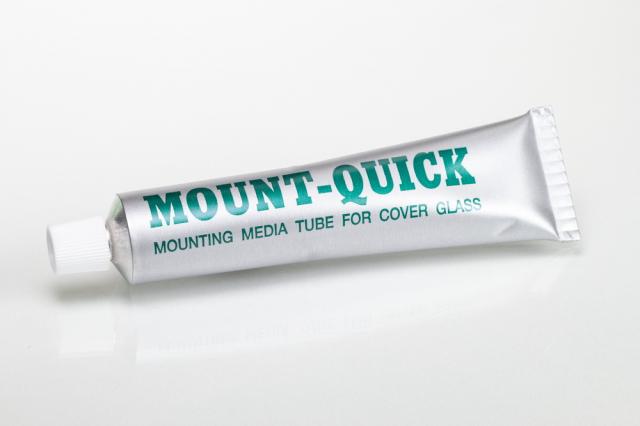
Mount-Quick
Tissue Transfer Procedure
-
- Xylene soluble mounting medium
- Tissue transfer medium
- Fast drying
- Clear
- 30cc tube packaging
- Manufactured by Daido Sangyo Co. Ltd.
![]()

PRODUCT:
| 30 cc Tube | |
| Mount-Quick Mounting Media | Part 6270 |
Additionally Needed for Tissue/Cell Transfer Procedure:
| Xylene, ACS | Part 1445 |
| HistoTec™ Pen | Part 5725 |
| Scalpels, Finger OR Scalpels, Flat Edge |
Part 6810 OR Part 5545 |
| Superfrost Plus Slides OR NewSilane Adhesive Slides |
Part 6203W OR Part 5070 |
| Alcohol, Ethyl Denatured, 100% | Part 10841 |
| Alcohol, Ethyl Denatured, 95% | Part 10842 |
APPLICATION:
Newcomer Supply Mount-Quick Mounting Media is a multi-use product with applications as a standard mounting media and as a liquid coverslip (coverglass free) mounting media.
Mount-Quick Mounting Media can also be used in tissue transfer procedures to transfer tissue sections or cell preparations from a prepared slide when specimen availability is limited, and additional diagnostic workup is necessary. Examples when tissue transfer with Mount-Quick Mounting Media is applicable include:
-
- Paraffin sections of small biopsies/minimal tissue
- Biopsies with a small foci of neoplasm
- Paraffin block is unavailable
- Cytology preparations, fine needle aspirates (FNA)
- Transfer of tissue from an uncharged slide to charged slide(s)
- Preparing multiple slides from a single slide preparation
- Salvaging/restoring broken slides back to a whole mount
This tissue/cell transfer procedure takes time to obtain a well-prepared end product. Reduced timings/short cuts should not be taken. Practicing the technique prior to performing on limited specimens is suggested. Staining quality of tissues will not be affected and histochemical, immunohistochemistry (IHC) and cytochemical procedures can be performed on transferred samples with reliable results.
METHOD:
Coverslipping: Paraffin or frozen sections
Tissue/Cell Transfer: Stained and unstained paraffin sections, cytology preparations and smears.
COVERSLIPPING PROCEDURE:
-
- Complete staining procedure: dehydrate, clear with xylene, blot excess xylene from slide edges.
- Apply Mount-Quick Mounting Media to flow over and fully cover the tissue section(s).
-
- Standard coverglass; install one edge first, allowing air bubbles to escape as opposite edge is lowered to slide.
- Liquid coverglass (coverglass free); allow to dry flat for 30 minutes before handling and viewing microscopically.
-
- Dry slides a minimum of 24-48 hours before filing.
TISSUE/CELL TRANSFER PROCEDURE:
-
- Mark designated tissue area(s) to be the focus of the tissue transfer on underside of the original slide with a HistoTec Pen.
- For previously stained sections:
-
- Soak slide in xylene until coverslip is easily removed without damage to tissue section.
- Run slide through several changes of xylene to ensure removal of residual mounting media.
- Do not proceed through alcohol steps.
- See Procedure Note #1.
-
- For unstained paraffin sections or smears:
-
- Soak slides in several changes of xylene, a minimum of 10 minutes. Do not proceed through alcohol steps.
-
- Spread Mount-Quick Mounting Media on xylene-coated slides with an applicator stick. Use sufficient Mount-Quick to form a meniscus over entire tissue section or cell preparation.
-
- Slide must be xylene coated and not allowed to dry out.
-
- Place slide in a flat horizontal position in a 60°C oven for 90 minutes to 2 hours or in a 37°C oven overnight until meniscus hardens.
-
- See Procedure Note #2.
-
- Mark the surface of the hardened mounting media meniscus to corresponding areas marked on underside of the slide (Step #1) with HistoTec Pen to maintain tissue orientation in transfer process.
- Immerse the slide in a Coplin jar of water warmed in a 45°- 60°C water bath; soak for 90 minutes to 3 hours.
- Slowly pry meniscus off at the edges with a scalpel blade. If the mounting media does not peel off easily, return to warm Coplin jar and continue soaking.
-
- See Procedure Note #3.
-
- Moisten the charged slides that will receive the transferred segments with water.
- Divide transferred segment into multiple sections: once media section is removed from the original slide, cut section with a water moistened scalpel blade into desired segments as marked on mounting media surface.
-
- Place each cut segment, meniscus side up, onto a moistened charged slide in sequential order.
-
- To transfer one intact segment of tissue: place on moistened charged slide, meniscus side up.
- Soak gauze in warm water, place gauze over tissue section. Using thumb, press gauze flat transferring water to the section, flattening edges and sealing transferred section to slide.
-
- If a transferred section is placed meniscus down, the specimen will not adhere and will be lost in further steps.
-
- Place slides in a 37°- 60°C oven in a flat horizontal position for 1 hour or longer to securely adhere transferred section to slide.
- Soak slides in four changes of xylene for 10 minutes each or longer until all Mount-Quick compound is completely dissolved.
-
- See Procedure Note #4.
-
- Re-hydrate through two changes each of 100% and 95% ethyl alcohols, 10 dips each. Wash well with distilled water.
- Specimen is ready for histochemical, IHC or cytochemical staining.
PROCEDURE NOTES:
-
- If any residual mounting medium remains on the slide, tissue transfer and staining may be compromised.
- The meniscus of Mount-Quick Mounting Media must thoroughly harden (but still remain pliable) on the slide before proceeding. To avoid brittleness do not exceed 60°C during the hardening process.
- If a hotplate was used to initially dry slides or melt paraffin after original sectioning, it will be difficult to remove an entire intact section from the slide. Extended soaking of mounting media and tissue section does not improve results.
- If Mount-Quick is not completely dissolved from the tissue section(s), subsequent staining may be compromised.
- The use of xylene substitutes has not been tested with this product.
REFERENCES:
-
- Gill, Gary. Cytopreparation: Principles & Practice, Essentials in Cytopathology. New York: Springer Science and Business Media, 2013. 393-395.
- Kubier, Patty and Rodney Miller. “Tissue Protection Immunohistochemistry.” American Journal of Clinical Pathology 117 (2002): 194-198.
- Miller, Rodney and Patty Kubier. “Immunohistochemistry on Cytologic Specimens and Previously Stained Slides (When No Paraffin Block is Available).” The Journal of Histotechnology4 (2002): 251-257.
- Sherman ME, D Jimenez-Joseph, MD Gangi and RR Rojas-Corona. “Immunostaining of Small Cytologic Specimens. Facilitation with Cell Transfer.” Acta Cytologica1 (1994): 18–22.
- Zu, Youli, Maryann Gangi, and Grace Yang, “Ultrafast Papanicolaou Stain and Cell-Transfer Technique Enhance Cytologic Diagnosis of Hodgkin Lymphoma.” Diagnostic Cytopathology 5 (2002): 308-311.
- Modifications developed by Newcomer Supply Laboratory.