Elastic, Verhoeff Stain Kit
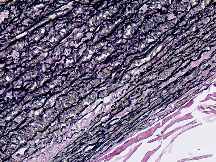
Used to demonstrate pathologic changes in elastic fibers as well as demonstration of normal elastic tissue such as arteries and veins.
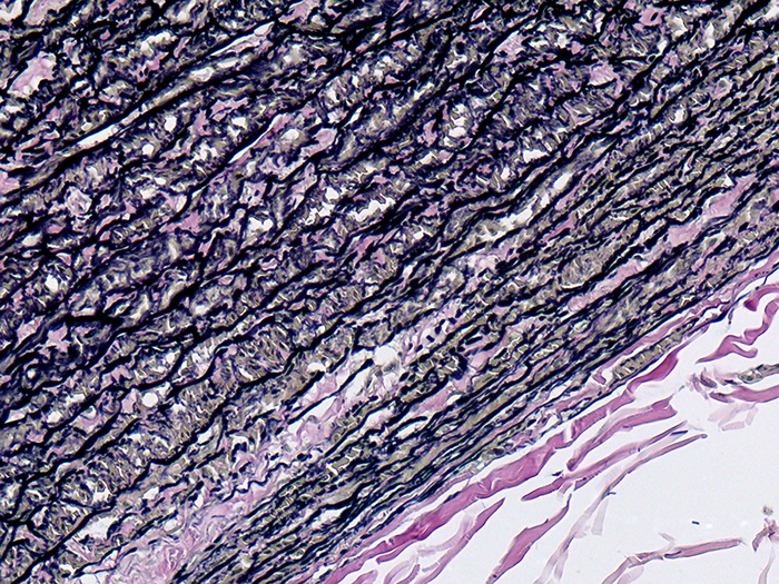
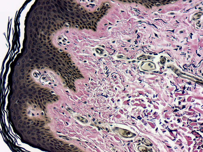
![]()


ELASTIC, VERHOEFF STAIN KIT INCLUDES:
| Part 9116A | Part 9116B | ||
| Solution A: | Hematoxylin 5%, Alcoholic | 125 ml | 250 ml |
| Solution B: | Ferric Chloride 10%, Aqueous | 125 ml | 250 ml |
| Solution C: | Iodine, Lugol’s, Aqueous | 75 ml | 150 ml |
| Solution D: | Sodium Thiosulfate 5%, Aqueous | 250 ml | 500 ml |
| Solution E: | Van Gieson Stain | 250 ml | 500 ml |
COMPLIMENTARY POSITIVE CONTROL SLIDES: Enclosed are two complimentary unstained positive control slides for the initial verification of staining techniques and reagents. Verification must be documented by running one Newcomer Supply complimentary positive control slide along with your current positive control slide for the first run. Retain the second complimentary control slide for further troubleshooting, if needed.
Individual stain solutions and additional control slides may be available for purchase under separate part numbers.
Additionally Needed:
| Xylene, ACS | Part 1445 |
| Alcohol, Ethyl Denatured, 100% | Part 10841 |
| Alcohol, Ethyl Denatured, 95% | Part 10842 |
For storage requirements and expiration date refer to individual bottle labels.
APPLICATION:
Newcomer Supply Elastic, Verhoeff Stain Kit procedure, commonly referred to as Verhoeff-Van Gieson (VVG) technique, is used to demonstrate pathologic changes in elastic fibers as well as demonstration of normal elastic tissue such as arteries and veins.
METHOD:
Fixation: Formalin 10%, Phosphate Buffered (Part 1090)
Technique: Paraffin sections cut at 4 microns
Solutions: All solutions are manufactured by Newcomer Supply, Inc.
All Newcomer Supply Stain Kits are designed to be used with Coplin jars filled to 40 ml following the provided staining procedure. Some solutions in the kit may contain extra volumes.
PRESTAINING PREPARATION:
-
- If necessary, heat dry tissue sections/slides in oven.
- Prepare fresh Verhoeff Working Solution by combining in the exact order listed, mixing well after each addition. Save for Step #5.
- Solution A: Hematoxylin 5%, Alcoholic 20 ml
- Solution B: Ferric Chloride 10%, Aqueous 8 ml
- Solution C: Iodine, Lugol’s, Aqueous 8 ml
- Prepare fresh Ferric Chloride 2%, Aqueous Solution for Step #7.
- Solution B: Ferric Chloride 10%, Aqueous 10 ml
- Distilled water 40 ml
STAINING PROCEDURE:
-
- Deparaffinize sections thoroughly in three changes of xylene, 3 minutes each. Hydrate through two changes each of 100% and 95% ethyl alcohols, 10 dips each. Wash well with distilled water.
- See Procedure Notes #1 and #2.
- Stain in fresh Verhoeff Working Solution (Step #2) for 30 minutes.
- Discard solution after successful differentiation in Step #7.
- Rinse in several changes of tap water.
- Differentiate each slide individually with agitation, in fresh Ferric Chloride 2%, Aqueous Solution (Step #3); approximately 5-10 dips.
- Check differentiation: rinse well in tap water and check microscopically for black elastic staining with gray background.
- If necessary, return to Ferric Chloride 2%, Aqueous Solution until desired elastic differentiation is achieved.
- See Procedure Notes #3 and #4.
- Deparaffinize sections thoroughly in three changes of xylene, 3 minutes each. Hydrate through two changes each of 100% and 95% ethyl alcohols, 10 dips each. Wash well with distilled water.
-
- Wash well in tap water.
- Place in Solution D: Sodium Thiosulfate 5%, Aqueous for 1 minute.
- Wash well in running tap water for 5 minutes.
- Counterstain in Solution E: Van Gieson Stain for 3 to 5 minutes.
- Dehydrate in two changes each of 95% and 100% ethyl alcohol. Clear in three changes of xylene, 10 dips each; coverslip with compatible mounting medium.
RESULTS:
| Elastic fibers/tissue | Blue-black to black |
| Collagen | Red |
| Other tissue elements | Yellow |
PROCEDURE NOTES:
-
- Drain slides after each step to prevent solution carry over.
- Do not allow sections to dry out at any point during procedure.
- If background is colorless, the section has been over-differentiated.
- Return to Step #5, restain and reduce differentiation dips.
- Differentiation will vary between slides depending on amount of elastic present in sections.
- If using a xylene substitute, closely follow the manufacturer’s recommendations for deparaffinization and clearing steps.
REFERENCES:
-
- Carson, Freida L., and Christa Hladik Cappellano. Histotechnology: A Self-instructional Text. 4th ed. Chicago: ASCP Press, 2015. 167-169.
- Mallory, Frank Burr, and James Homer Wright. Pathological Technique. 7th ed. Philadelphia, PA: W.B. Saunders Company, 1918. 118-119.
- Modifications developed by Newcomer Supply Laboratory.