
Urates, Gomori Methenamine-Silver
Reagents for this procedure are sold as individual stain solutions and are available for purchase under separate part numbers with storage requirements and expiration date designated per bottle.
![]()
SOLUTIONS:
| 100 ml | 250 ml | 500 ml | |
| Methenamine 3%, Aqueous | Part 12239A | Part 12239B | |
| Silver Nitrate 5%, Aqueous | Part 13805A | Part 13805B | |
| Sodium Borate 5%, Aqueous | Part 13826B | ||
| Gold Chloride 0.25%, Aqueous | Part 11287A | Part 11287B | |
| Sodium Thiosulfate 2.5%, Aqueous | Part 13889A | Part 13889B | |
| Light Green SF Yellowish Stain 0.2%, Aqueous | Part 12202A | Part 12202B |
Additionally Needed:
| Urates Control Slides | Part 4700 |
| Hydrochloric Acid 5%, Aqueous | Part 12086 (for acid cleaning glassware) |
| Xylene, ACS | Part 1445 |
| Alcohol, Ethyl Denatured, 100% | Part 10841 |
| Alcohol, Ethyl Denatured, 95% | Part 10842 |
For storage requirements and expiration date refer to individual product labels.
APPLICATION:
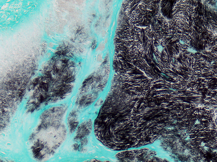
Newcomer Supply Urate Stain, Gomori Methenamine Silver Method is designed to demonstrate urates in tissue sections. With abnormal accumulations found around joints and in soft tissues, this disturbance in uric acid metabolism is known as gout, with collections of urate crystals referred to as gouty tophi.
Calcium pyrophosphate crystals or pseudogout may also be demonstrated. When viewed with a polarizing filter and red compensator filter, gout and pseudogout can be distinguished.
METHOD:
Fixation: Urate crystals are soluble in aqueous solutions. Fix in 100% ethyl alcohol; a minimum of two changes, 4 hours each.
Processing: Transfer from 100% ethyl alcohol fixative to xylene for 1 hour; proceed with equal parts xylene/paraffin at 58°C for 2 hours. Infiltrate with paraffin for a minimum of 1 hour; embed.
Technique: Chill paraffin blocks in 100% ethyl alcohol; cut sections at 4 microns with minimal water bath exposure.
Solutions: All solutions are manufactured by Newcomer Supply, Inc.
All Newcomer Supply stain procedures are designed to be used with Coplin jars filled to 40 ml following the provided staining procedure.
PRESTAINING PREPARATION:
-
- If necessary, heat dry tissue sections/slides in oven.
- All glassware/plasticware must be acid cleaned prior to use.
-
- See Procedure Notes #1 and #2.
-
- Prepare Methenamine Silver Stock Solution.
-
- Methenamine 3%, Aqueous (Part 12239) 50 ml
- Silver Nitrate 5%, Aqueous (Part 13805) 5 ml
- Slowly add silver nitrate; mix to clear milky precipitate.
- Store clear stock solution at 2°C-8°C for up to 2 months.
-
- Prepare fresh Methenamine Silver Working Solution; mix well.
-
- Methenamine Silver Stock Solution 25 ml
- Distilled Water 25 ml
- Sodium Borate 5%, Aqueous (Part 13826) 3 ml
-
- Preheat fresh Methenamine Silver Working Solution to 60°C in a water bath.
STAINING PROCEDURE:
-
- Deparaffinize sections thoroughly in three changes of xylene, 3 minutes each. Rinse in two changes of 100% ethyl alcohol, 10 dips each.
-
- Do not use 95% ethyl alcohol or distilled water steps.
- See Procedure Notes #3 and #4.
-
- Deparaffinize sections thoroughly in three changes of xylene, 3 minutes each. Rinse in two changes of 100% ethyl alcohol, 10 dips each.
-
- Incubate slides in preheated Methenamine Silver Working Solution in a 60°C water bath for 30 minutes.
-
- Remove control slide, rinse in warm distilled water, check microscopically for adequate silver development. Crystals should be dark brown/black.
- If not sufficiently dark, return to warm silver solution.
- Recheck at 2-3-minute intervals for desired intensity.
-
- Rinse well in distilled water.
- Tone in Gold Chloride 0.25%, Aqueous (Part 11287) until brown colorization disappears; 5 to 30 seconds.
- Rinse well in distilled water.
- Place in Sodium Thiosulfate 2.5%, Aqueous (Part 13889); 2-3 minutes.
- Wash well in running tap water for 3 minutes; rinse in distilled water.
- Counterstain in Light Green SF Yellowish Stain 0.2%, Aqueous (Part 12202) for 1-2 minutes.
- Dehydrate in two changes each of 95% and 100% ethyl alcohol. Clear in three changes of xylene, 10 dips each; coverslip with compatible mounting medium.
- Incubate slides in preheated Methenamine Silver Working Solution in a 60°C water bath for 30 minutes.
RESULTS:
| Light Field Microscopy: | ||
| Gout/urate crystals | Black | |
| Background | Green | |
| Polarized/Red Compensator Filter: (long axes aligned parallel) | ||
| Gout/urate crystals | Yellow, long & needle shaped | |
| Pseudogout crystals | Blue, shorter & rhomboidal | |
PROCEDURE NOTES:
-
- Acid clean all glassware/plasticware (Part 12086) and rinse thoroughly in several changes of distilled water.
- Plastic (Part 5500), plastic-tipped, or paraffin coated metal forceps must be used with any silver solution to prevent precipitation of silver salts. No metals of any kind should be in contact with any silver solution. Only glass thermometers should be used.
- Drain slides after each step to prevent solution carry over.
- Do not allow sections to dry out at any point during procedure.
- If using a xylene substitute, closely follow the manufacturer’s recommendations for deparaffinization and clearing steps.
REFERENCES:
-
- Carson, Freida L., and Christa Hladik. Histotechnology: A Self-Instructional Text. 3rd ed. Chicago, Ill.: American Society of Clinical Pathologists, 2009.255-256, 267-268.
- Luna, Lee G. Manual of Histologic Staining Methods of the Armed Forces Institute of Pathology. 3rd ed. New York: Blakiston Division, McGraw-Hill, 1968. 187-188.
- Sheehan, Dezna C., and Barbara B Hrapchak. Theory and Practice of Histotechnology. 2nd ed. St. Louis: Mosby, 1980. 225-226.
- Modifications developed by Newcomer Supply Laboratory.