Fungus, PAS/Light Green Stain Kit
Stains fungus and glycoproteins. Polysaccharides present in fungal cell walls are oxidized by the periodic acid to aldehydes. The aldehydes then react with Schiff Reagent, McManus to yield magenta colored fungi.
![]()

FUNGUS, PAS/LIGHT GREEN STAIN KIT INCLUDES:
| Part 9122A | ||
| Solution A: | Periodic Acid 0.5%, Aqueous | 250 ml |
| Solution B: | Schiff Reagent, McManus | 250 ml |
| Solution C: | Light Green SF Yellowish Stain 0.1%, Aqueous | 250 ml |
COMPLIMENTARY POSITIVE CONTROL SLIDES: Enclosed are two complimentary unstained positive control slides for the initial verification of staining techniques and reagents. Verification must be documented by running one Newcomer Supply complimentary positive control slide along with your current positive control slide for the first run. Retain the second complimentary control slide for further troubleshooting, if needed.
Individual stain solutions and additional control slides may be available for purchase under separate part numbers.
Additionally Needed:
| Xylene, ACS | Part 1445 |
| Alcohol, Ethyl Denatured, 100% | Part 10841 |
| Alcohol, Ethyl Denatured, 95% | Part 10842 |
For storage requirements and expiration date refer to individual bottle labels.
APPLICATION:
Newcomer Supply Fungus, PAS/Light Green Stain Kit procedure is used for identifying fungus and glycoproteins in tissue sections. Polysaccharides present in fungal cell walls are oxidized by periodic acid to aldehydes; aldehydes then react with Schiff Reagent, McManus to yield magenta colored fungi.
METHOD:
Fixation: Formalin 10%, Phosphate Buffered (Part 1090)
Technique: Paraffin sections cut at 4 microns
Solutions: All solutions are manufactured by Newcomer Supply, Inc.
All Newcomer Supply Stain Kits are designed to be used with Coplin jars filled to 40 ml following the staining procedure provided below. Some solutions in the kit may contain extra volumes.
STAINING PROCEDURE:
-
- If necessary, heat dry tissue sections/slides in oven.
- Deparaffinize sections thoroughly in three changes of xylene, 3 minutes each. Hydrate through two changes each of 100% and 95% ethyl alcohols, 10 dips each. Wash well with distilled water.
-
- See Procedure Notes #1 and #2.
-
- Place in Solution A: Periodic Acid 0.5%, Aqueous for 5 minutes.
- Wash in three changes of tap water; rinse in distilled water.
- Drain slides of excess water and stain in Solution B: Schiff Reagent, McManus for 20 minutes.
- Wash gently in lukewarm tap water for 10 minutes to allow pink color to develop.
- Counterstain in Solution C: Light Green SF Yellowish Stain 0.1%, Aqueous for 5 seconds.
-
- See Procedure Note #3.
-
- Dehydrate in two changes each of 95% and 100% ethyl alcohol. Clear in three changes of xylene, 10 dips each; coverslip with compatible mounting medium.
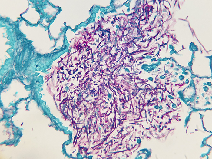
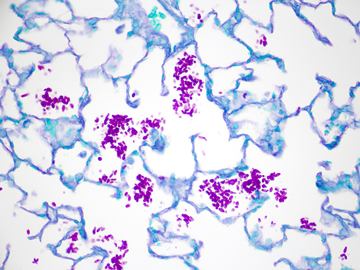
RESULTS:
| Fungal cell walls and glycogen | Red to magenta |
| Background | Pale green |
PROCEDURE NOTES:
-
- Drain slides after each step to prevent solution carry over.
- Do not allow sections to dry out at any point during procedure.
- Increase or decrease staining time in Solution C: Light Green SF Yellowish Stain 0.1%, Aqueous for preference of counterstain intensity.
- If using a xylene substitute, closely follow the manufacturer’s recommendations for deparaffinization and clearing steps.
REFERENCES:
-
- Bancroft, John D., and Marilyn Gamble. Theory and Practice of Histological Techniques. 6th ed. Oxford: Churchill Livingstone Elsevier, 2008. 321-323.
- Sheehan, Dezna C., and Barbara B. Hrapchak. Theory and Practice of Histotechnology. 2nd ed. St. Louis: Mosby, 1980. 245.
- Modifications developed by Newcomer Supply Laboratory.